

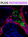
Confocal Microscopy of Reovirus Transport in Living Dorsal Root Ganglion Neurons
活体背根节神经元中呼肠孤病毒转运的共聚焦显微镜观察
发布: 2020年11月20日第10卷第22期 DOI: 10.21769/BioProtoc.3825 浏览次数: 4467
评审: John SL ParkerAbraam YakoubKristin L. Shingler
参见作者原研究论文
The authors used this protocol in:
Feb 2020
Advertisement


Abstract
Neurotropic reoviruses repurpose host machinery to traffic over long distances in neuronal processes and access distal replication sites. Understanding mechanisms of neuronal transmission is facilitated by using simplified in vitro primary neuronal culture models. Advances in the design of compartmentalized microfluidic devices lend robustness to neuronal culture models by enabling compartmentalization and manipulation of distinct neuronal processes. Here, we describe a streamlined methodology to culture sensory neurons dissociated from dorsal root ganglia of embryonic rats in microfluidic devices. We further describe protocols to exogenously label reovirus and image, track, and analyze transport of single reovirus particles in living neurons. These techniques can be adapted to study directed axonal transport of other neurotropic viruses and neuronal factors involved in signaling and pathology.
Background
Viruses from diverse families including the Flaviviridae, Herpesviridae, Picornaviridae, and Rhabdoviridae, breach protective barriers of the nervous system inflicting severe disease and economic burden (Koyuncu et al., 2013; Bohmwald et al., 2018; Tyler, 2018). Mammalian orthoreovirus (reovirus), belonging to the Reoviridae, causes serotype-dependent neuronal infection in a wide variety of young mammals that can result in lethal encephalitis (Tyler et al., 1986; Dermody et al., 2013]. Reovirus is nonenveloped with a segmented dsRNA genome encased by two concentric protein shells and serves as a pliable tool to study viral infections of the nervous system (Dermody et al., 2013). Although cellular and molecular mechanisms of reovirus infection have been widely studied using transformed cell lines, these systems do not capture the complexities of polarized neuronal cells. Viruses infecting neurons must travel over long distances in axons to reach distal sites of replication and egress. To understand mechanisms of reovirus entry and long-distance transport in neurons, we recently adapted techniques to culture primary neurons and image fluorescently labeled reovirus in living cells (Aravamudhan et al., 2020).
In vitro culture of primary neurons from embryonic and adult rodents provide simplified yet robust tools to study neuronal function and pathology. Dissociated sensory neurons from dorsal root ganglia (DRG) can be propagated in vitro and are used widely to study axonal regeneration and peripheral neuropathies (Haberberger et al., 2019; Melli and Hoke, 2009). DRGs are clusters of neuronal cell bodies located proximal to the spinal cord and relay sensory information from the periphery to the central nervous system. DRG neurons are pseudo-unipolar with no dendrites and one axon that bifurcates into two processes, one synapsing with peripheral organs and another connecting to the spinal cord. DRGs are infected during neuroinvasion by several viruses and have been used to study infection mechanisms (Flamand et al., 1991; Smith et al., 2001; Volpi et al., 2018). We describe procedures to cultivate dissociated DRG neurons from rat embryos. We also have used this protocol to cultivate DRG neurons from embryonic mice, which provide a more robust system amenable to genetic manipulation. However, we found that reovirus infects rat neurons more efficiently than mouse neurons in culture (unpublished observations). Therefore, the system must be chosen carefully depending on the application.
In vitro neuron culture in combination with microfluidic devices offers a powerful system to isolate neuronal soma from axons and distinguish axonal transport in the anterograde (soma to axonal termini) and retrograde (axonal termini to soma) directions (Neto et al., 2016). Microfluidic devices allow use of small quantities of reagents and provide access to neuronal microenvironments for studies of axonal growth and injury, biochemical investigations, and high-resolution imaging. The commercial availability of microfluidic devices makes it accessible to everyone without need of sophisticated fabrication instruments. We describe use of a commercially available two-compartment system for culturing DRG neurons and imaging reovirus transport (Nagendran et al., 2018). This device was developed by Xona microfluidics and comes preassembled on optically transparent plastic suitable for high-resolution imaging.
We share an integrated set of protocols to isolate and culture DRG neurons in microfluidic devices to study reovirus transport. In addition, we describe image analysis tools to track single reovirus particles and share programs to obtain biophysical parameters describing reovirus transport. These methods can be adapted to investigate molecular mechanisms of viral neuronal infection. Individual aspects of this procedure can be applied to fluorescently label other nonenveloped viruses, study transport of other neurotropic pathogens, and dissect functions of local RNA and protein metabolism in axonal signaling. Extension of co-culture techniques can be used to study communication between neurons and glia and peripheral tissues. Culture of neurons in microfluidic devices embedded with electrode arrays can be used to simultaneously study electrical activity and biochemical signaling, further enhancing the utility of protocols described here.
Materials and Reagents
Sterilization pouches (VWR, catalog number: 58753-194 )
Transfer pipette (VWR, catalog number: 414004-002 )
Glass Pasteur pipette (VWR, catalog number: 14673-043 )
Plug the wide end with of the glass pipette with cotton. Polish the capillary end using a Bunsen burner by holding the tip in the flame and rotating it rapidly. Pause every few seconds and check the size of the opening to ensure that the edge is smooth and round and the opening is not too narrow. A narrow opening will disrupt cells during the trituration. Autoclave the fire polished Pasteur pipette to sterilize before use.
35 mm Petri dishes (VWR, Greiner Bio-One, catalog number: 82050-540 )
100 mm Petri dishes (VWR, catalog number: 25384-342 )
150 mm Petri dishes (Midland Scientific Inc., Kord Valmark, catalog number: VM 2902 )
Microfluidic devices (Xona microfluidics, catalog number: XC450 )
Glass bottom dishes (MatTek, catalog number: P35G-1.5-14-C )
Closures for dialysis membrane tubing (VWR, Spectrum Laboratories [132735], catalog number: 25224-075 )
Glass vial (Qorpak, catalog number: GLC-00987 )
Dialysis membrane tubing, MWCO 12000-14000 (Fisher Scientific, SpectrumTM S432697, catalog number: 08-667A )
Syringe filter 0.22 μm (VWR, catalog number: 28145-477 )
Conical tubes (VWR, Corning, catalog number: 21008-656 )
Eppendorf tubes (FisherbrandTM, catalog number: 0 5408129 )
E14.5 pregnant Sprague Dawley rat
Immersion oil for microscopy (Cargille, catalog number: 16484 )
Papain (Worthington Biochemical, catalog number: LS003126 )
L-cysteine (Sigma-Aldrich, catalog number: C7352 )
Ovomucoid protease inhibitor (Worthington, catalog number: LK003182 ). Reconstitute in 32 ml of HBSS and store at 4 °C
Collagenase A (Roche, catalog number: 10103578001 )
DNase I (Sigma-Aldrich, Catalog number: D5025-15KU )
Trypan blue (Sigma-Aldrich, catalog number: T8154 )
Hanks’ balanced salt solution without calcium and magnesium, HBSS (CellGro-Corning, catalog number: 21-022-CV )
Poly-D-lysine hydrobromide, PDL (Sigma-Aldrich, catalog number: P0899 ). Reconstitute in HBSS at a concentration of 0.5 mg/ml, filter sterilize, make 5 ml aliquots in sterile conical tubes and store at -20 °C
Laminin (Corning, catalog number: 354232 )
Thaw slowly on ice, make aliquots of different volumes (10 to 50 μl) in sterile tubes, and store at -20 °C. Laminin will form a gel following rapid changes in temperature. For coating, combine multiple aliquots of different volumes to arrive at the final required volume. Do not re-freeze after thawing.
Alexa FluorTM 647 NHS Ester (Thermo Fisher Scientific, catalog number: A37573 ). Reconstitute at 10 mM concentration in DMSO using sterile conditions and make 2 μl aliquots. Store at -20 °C and use within 6 months. The dye solution becomes hydrolyzed over time, resulting in poor labeling efficiency. Do not refreeze after thawing.
Sodium bicarbonate, NaHCO3 (Fisher chemicalTM, catalog number: S233-500 )
NeurobasalTM medium (Gibco, catalog number: 21103049 )
UltraCULTURETM serum-free (Lonza, catalog number: BE12-725F )
GlutaMAXTM supplement (Gibco, catalog number: 35050079 )
Penicillin-streptomycin (Gibco, catalog number: 15070-063 )
HyCloneTM fetal bovine serum (GE Healthcare/Cytiva, catalog number: SH30088.03 ). Make 50 ml aliquots in sterile conical tubes and store at -20 °C. Store thawed aliquots at 4 °C up to 6 months
N-2 supplement (Gibco, catalog number: 17502048 ). Thaw on ice, make 500 μl aliquots in sterile tubes, and store at -20 °C
B-27TM supplement (Gibco, catalog number: 17504044 ). Thaw on ice, make 1 ml aliquots in sterile tubes, and store at -20 °C
Nerve growth factor 2.5S subunit, NGF (Gibco, catalog number: 13257019 ). Thaw on ice, make 15 μl aliquots in sterile tubes, and store at -20 °C. Supplied at 100 µg/ml
Hibernate-E medium (Gibco, catalog number: A1247601 )
Cytosine β-D-arabinofuranoside, AraC (Sigma-Aldrich, catalog number: C1768 ). Reconstitute in water at a concentration of 10 mM, filter sterilize, make 100 μl aliquots in sterile tubes, and store at -20 °C. To limit repeated freeze-thaws, each aliquot can be re-aliquoted in 10 μl upon thawing and stored at -20 °C
NaCl (Fisher ChemicalTM, catalog number: S671-3 )
KCl (Fisher ChemicalTM, catalog number: P217-500 )
Na2HPO4 (Sigma-Aldrich, catalog number: S-7907 )
KH2PO4 (Fisher ChemicalTM, catalog number: P285-500 )
Collagenase-DNase I mixture (see Recipes)
Base medium (see Recipes)
Complete medium (see Recipes)
Low NGF medium (see Recipes)
Papain solution (see Recipes)
10x PBS (see Recipes)
10x sodium bicarbonate buffer (see Recipes)
Equipment
Scissors (Fine Science Tools, catalog number: 91460-11 )
Forceps (2) (Fine Science Tools, catalog number: 11253-20 )
Spring scissors (Fine Science Tools, catalog number: 15023-10 )
Sharp forceps (Fine Science Tools, Dumont #5, catalog number: 11252-20 )
Dissection microscope (CarlZeissTM, StemiTM DV4 )
Spectrophotometer (Thermo Fisher Scientific, model: NanoDropTM 1000 )
Tube rotator (VWR, catalog number: 10136-084 )
Tabletop centrifuge (Thermo ScientificTM, model: SorvallTM LegendTM Micro 17R )
pH meter (VWR, model: SympHonyTM B10P )
Tissue culture (TC) hood (BSL-2 certified)
TC microscope (ZeissTM, model: InvertoskopTM 40C )
Hemacytometer (Bright-LineTM, Millipore sigma, catalog number: Z359629 )
Software
Fiji ImageJ (https://fiji.sc/; Rasband, 1997-2018; Schneider et al., 2012 )
MATLAB (version 9.07.0 [R2019b]; Natick, Massachusetts: The MathWorks Inc. 2019; https://www.mathworks.com/products/matlab.html)
Procedure
文章信息
版权信息
© 2020 The Authors; exclusive licensee Bio-protocol LLC.
如何引用
Aravamudhan, P., Raghunathan, K. and Dermody, T. S. (2020). Confocal Microscopy of Reovirus Transport in Living Dorsal Root Ganglion Neurons. Bio-protocol 10(22): e3825. DOI: 10.21769/BioProtoc.3825.
分类
神经科学 > 周围神经系统
微生物学 > 微生物细胞生物学
细胞生物学 > 基于细胞的分析方法
您对这篇实验方法有问题吗?
在此处发布您的问题,我们将邀请本文作者来回答。同时,我们会将您的问题发布到Bio-protocol Exchange,以便寻求社区成员的帮助。