

Cleavable Affinity Purification (Cl-AP): A One-step Procedure to Affinity Purify Protein Complexes
CI-AP:一种亲和纯化蛋白质复合物的一步法
发布: 2020年11月20日第10卷第22期 DOI: 10.21769/BioProtoc.3821 浏览次数: 4604
评审: Abhijit KaleSandhya GanesanAnonymous reviewer(s)
参见作者原研究论文
The authors used this protocol in:

Jan 2020
Advertisement


Abstract
Cleavable Affinity Purification (Cl-AP) uses a tripartite system of Protein-A-Streptavidin beads and nanobodies, coupled with a biotinylated, thiol-cleavable linker, providing one-step affinity purification from lysates of tissues expressing tagged proteins. This technique allows fluorescent versions of mitotic protein complexes to be isolated intact from cells, for use in biophysical and microscopy-based assays, overcoming the traditional limitations of reductionist approaches. We have used this technique successfully to purify both GFP-tagged and mCherry-tagged proteins, and their interacting partners, expressed in Drosophila melanogaster embryos. Although we demonstrate the efficacy of the GFP-binding protein and RFP-binding protein nanobodies from Chromotek, in theory any antibody could be coupled to the beads and used as a Cl-AP reagent.
Keywords: Drosophila melanogaster (黑腹果蝇)Background
Many proteins elicit their cellular function as part of multi-protein complexes. In order to gain full understanding of the role of a protein complex, in vivo approaches such as perturbation of protein levels/activity or monitoring dynamic localization need to be combined with in vitro biochemical assays and functional reconstitution. This holistic approach is currently severely limited. In vitro studies generally use proteins that have been individually expressed and purified in non-autogenous systems such as bacteria and insect cells, where folding and post translational modifications crucial to function may be dissimilar to that found in the original cell. Conversely, purification of specific proteins or complexes from cells/tissues generally relies on either co-immuno-precipitation or purification of a tagged version of the protein of interest which has been introduced into cells, for example using haemagglutinin (HA), FLAG3 or tandem affinity purification (TAP)-tagging. There are two main issues with these in vivo approaches: (i) the beads themselves bind to non-specific proteins leading to contamination problems, (ii) the strong antibody-antigen or tag-matrix interaction cannot be disrupted without disrupting the interactions between protein complex subunits. Modified protocols, in which the antibody-antigen interactions are disrupted using peptide competition or in which the tag is cleaved using proteases can potentially result in the purification of soluble, intact protein complex from cells. However, the former requires extensive knowledge of the antibody-antigen interface while both procedures are undertaken in non-physiological conditions and result in protein complex that requires further purification (separating the excess peptide or the protease respectively), complicating this type of approach.
We sought to develop a quick, easy and widely applicable technique that would solve these problems–allowing the isolation of a protein complex of interest in soluble form, in the buffer of choice. We chose GFP as our affinity tag, given the added advantage of being able to assess the dynamic localization and functionality of the fusion protein within a cell or tissue, prior to isolation. We term this technique Cleavable Affinity Purification (Cl-AP).
Materials and Reagents
15 ml conical tube
Beckman tube (Beckman Coulter, catalog number: 343776 )
Pipette tip with a diameter of 0.6 mm (e.g., Gel-Saver® II, STARLAB, catalog number: I1022-0600 )
Spin Desalting Column, 2 ml, 10 columns, for 200-700 μl samples, 7,000 MWCO (ZebaTM, catalog number: 89882 )
Tissue of choice (e.g., Drosophila embryos)
GFP VHH, recombinant binding protein (Chromotek, catalog number: gt-250 )
1x Phosphate-buffered Saline (PBS): 0.1 M sodium phosphate, 0.15 M NaCl, pH 7.2
Sulfo-NHS-SS-Biotin (Thermo Scientific, catalog number: 21945 )
Anhydrous dimethylformamide (DMF) (Sigma-Aldrich, catalog number: 227056 )
Buffer of choice (e.g., 1x BRB80: 80 mM PIPES, 1 mM EGTA, 1 mM MgCl2, pH 6.9 at 4 °C)
IGEPAL (Sigma, catalog number: I8896 )
Phenylmethylsulfonyl fluoride (PMSF) (Sigma, catalog number: 10837091001/PMSF-RO )
Pi (cOmplete Mini protease inhibitor) (Roche, catalog number: 11836170001 )Working concentration: 1 tablet per 10 ml extraction solution
PPi (PhosSTOP phosphatase inhibitor) (Roche, catalog number: 4906837001 )Working concentration: 1 tablet per 10 ml extraction solution
High Capacity Streptavidin Agarose Resin (Pierce, catalog number: 20349 )
Dithiothreitol (DTT) (Thermo scientific, catalog number: R0861 )
Biotinylated antibody/nanobody (Chromotek, catalog number: gt-250 )
Liquid nitrogen
Equipment
Optima MAX-TL ultracentrifuge with TLA-120.1 rotor (Beckman Coulter)
Microcentrifuge
Rocking platform shaker
Rotating disc mixer
Dounce homogenizer
-80 °C freezer
Procedure
Preparation of GFP-TRAP-Sulfo beads
Desalt/Buffer Exchange of the nanobody (e.g., GFP-binding-protein)
Note: The nanobody must be completely desalted into 1x PBS before being modified with Sulfo-NHS-SS-Biotin.
ZebaTM Spin desalting columns are used according to manufacturer's instructions.
Remove column’s bottom closure, loosen the cap, and place into a 15 ml conical tube.
Centrifuge at 1,000 x g for 2 min in a benchtop microfuge. Discard the flow-through (to remove storage buffer).
Place a mark on the side of the column where the compacted resin is slanted upward. Place column in the microcentrifuge with the mark facing outward in all subsequent centrifugation steps.
Equilibrate the Zeba column with 1 ml 1x PBS. Centrifuge at 1,000 x g for 2 min and discard the flow-through.
Repeat Step A4 two more times.
Insert the equilibrated column in a new conical tube. Apply 200 μl of nanobody (1 mg/ml) directly on the filter.
Centrifuge at 1,000 x g for 2 min and collect the flow through (nanobody).
Biotin-labelling Reaction
Allow vial of Sulfo-NHS-SS-Biotin to equilibrate to ambient temperature before opening.
Immediately before use, dissolve the Sulfo-NHS-SS-Biotin in DMF to give a 8 mM solution.
Add 86.1 μl of Sulfo-NHS-SS-Biotin solution to 200 μl of nanobody (1 mg/ml).
Mix immediately by inversion.
Incubate on ice for 2 h.
Stop the reaction by removing the excess biotin reagent using a fresh Zeba spin column (i.e., repeat Steps A1-A7).
Linking the Biotinylated antibody to Streptavidin Agarose beads
Mix the streptavidin agarose resin to ensure an even suspension.
Wash 40 μl of bead slurry (20 μl packed beads) by resuspension in 1x PBS, pelleting the beads at 2,500 x g for 2 min in a microcentrifuge. Repeat three more times.
Add biotinylated nanobody solution (from B6) to washed streptavidin agarose beads and allow to bind to beads using either a rocking platform shaker or a rotating disc mixer for 1 h at 4 °C.
Remove any unbound antibody by washing three times as described above (C2) but using the buffer of choice (e.g., BRB80 + 0.1% IGEPAL) instead of PBS.
After removing the supernatant from the last wash, resuspend the beads in the buffer of choice, to maintain a 50% (40 μl) bead slurry. Store at 4 °C.
Note: Cl-AP beads can be stored at 4 °C for at least 3 months with no loss of activity.
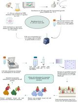
Homogenization of tissue and preparation of the High-speed Supernatant (HSS)
Drosophila embryos expressing the GFP-fusion protein of interest are collected (batches of 0- to 3-h-old embryos), dechorionated, weighed, flash frozen in liquid nitrogen and stored at -80 °C, until approximately 200 mg has been collected. All subsequent steps are performed on ice or in a 4 °C cold room using prechilled buffers and pipettes.
Note: Alternative tissues, for example tissue culture cells transiently expressing GFP- or mCherry-expressing proteins, can be substituted.
Transfer the tissue to a Dounce homogenizer on ice.
Add 400 μl of an appropriate homogenising buffer (e.g., BRB80 + 0.1% IGEPAL + PMSF + Pi and PPi) and homogenize.
Centrifuge homogenate at 14,000 x g for 10 min in a microfuge at 4 °C.
Transfer supernatant to a Beckman tube and spin at 100,000 x g for 10 min at 4 °C.
Transfer supernatant to a fresh tube and spin again for 30 min at 100,000 x g.
Incubation of HSS with cl-AP
Transfer the HSS to a new 1.5 ml tube and incubate with 20 μl (1:1 slurry) of GFP-TRAP-Sulfo beads, overnight in cold room on rotating wheel.
Cleaving of Sulfo-beads
Wash beads with assay buffer (e.g., 1 ml BRB80 + 0.1% IGEPAL) 4 times (Step C2) to remove unbound and non-specifically bound proteins.
After removing the supernatant from the last wash, add 50 μl of assay buffer containing 50 mM DTT (final concentration), to cleave the disulfide bond in the spacer arm.
Incubate sample in orbital shaker at RT for 2 h.
Pellet the beads at 2,500 x g for 2 min in a microcentrifuge.
Transfer the supernatant (eluate) to a fresh tube using a Gel-Saver II pipette tip.
Acknowledgments
This work was funded by University of Exeter (Proof of Concept), Lucy Green; University of Exeter (Carlota Palmer PhD studentship) Ammarah Tariq.
We thank Stephen Green and Mark Wood (Exeter, UK) for initial discussions about photocleavable and other tags, and Mary Munson (Massachusetts, US) for highlighting the potential of thiol-cleavable tags for native complex purification, as published by the Rout lab (Rockefeller, US) (Fridy et al., 2015). We thank Chromotek for the gift of GFP-binding protein; Jack Chen, Dan Hayward and Chris Sullivan for initial attempts to develop the Cl-AP technique.
The original research article where this protocol was used is Tariq et al., 2020.
Competing interests
No competing interests declared.
References
- Fridy, P.C., Thompson, M.K., Ketaren, N.E. and Rout, M.P. (2015). Engineered high-affinity nanobodies recognizing staphylococcal Protein A and suitable for native isolation of protein complexes. Anal Biochem 15: 477:92-4.
- Tariq, A., Green, L., Jeynes, J. C. G., Soeller, C. and Wakefield, J. G. (2020). In vitro reconstitution of branching microtubule nucleation. Elife 9: e49769.
文章信息
版权信息
![]() Tariq et al. This article is distributed under the terms of the Creative Commons Attribution License (CC BY 4.0).
Tariq et al. This article is distributed under the terms of the Creative Commons Attribution License (CC BY 4.0).
如何引用
Readers should cite both the Bio-protocol article and the original research article where this protocol was used:
- Tariq, A., Green, L., Soeller, C. and Wakefield, J. (2020). Cleavable Affinity Purification (Cl-AP): A One-step Procedure to Affinity Purify Protein Complexes. Bio-protocol 10(22): e3821. DOI: 10.21769/BioProtoc.3821.
- Tariq, A., Green, L., Jeynes, J. C. G., Soeller, C. and Wakefield, J. G. (2020). In vitro reconstitution of branching microtubule nucleation. Elife 9: e49769.
分类
生物化学 > 蛋白质 > 分离和纯化
您对这篇实验方法有问题吗?
在此处发布您的问题,我们将邀请本文作者来回答。同时,我们会将您的问题发布到Bio-protocol Exchange,以便寻求社区成员的帮助。