

Affinity Purification of GO-Matryoshka Biosensors from E. coli for Quantitative Ratiometric Fluorescence Analyses
用于定量比率荧光分析的大肠杆菌GO-Matryoshka生物传感器亲和纯化

发布: 2020年10月05日第10卷第19期 DOI: 10.21769/BioProtoc.3773 浏览次数: 4736
评审: Wolf Dieter RötherJing LiYue (Yolanda) HuangAnonymous reviewer(s)
参见作者原研究论文
The authors used this protocol in:

Sep 2017
Advertisement


Abstract
Genetically encoded biosensors are powerful tools for quantitative visualization of ions and metabolites in vivo. Design and optimization of such biosensors typically require analyses of large numbers of variants. Sensor properties determined in vitro such as substrate specificity, affinity, response range, dynamic range, and signal-to-noise ratio are important for evaluating in vivo data. This protocol provides a robust methodology for in vitro binding assays of newly designed sensors. Here we present a detailed protocol for purification and in vitro characterization of genetically encoded sensors, exemplified for the His affinity-tagged GO-(Green-Orange) MatryoshCaMP6s calcium sensor. GO-Matryoshka sensors are based on single-step insertion of a cassette containing two nested fluorescent proteins, circularly permutated fluorescent green FP (cpGFP) and Large Stoke Shift LSSmOrange, within the binding protein of interest, producing ratiometric sensors that exploit the analyte-triggered change in fluorescence of a cpGFP.
Keywords: Biosensors (生物传感器)Background
The green fluorescent protein (GFP) was identified in 1962 in the jellyfish Aequorea Victoria (Shimomura et al., 1962). Thirty years later, its first use as a reporter gene was described (Chalfie et al., 1994). Since their discovery, GFP variants and other fluorescent proteins have contributed greatly to the principal advancements in the biological sciences, and are now common tools in biomedical research (Frommer et al., 2009).
A large number of fluorescent proteins (FP) and FP variants have been used as reporters or fused to proteins in organisms of all kingdoms of life (Chudakov et al., 2010; Valeur and Berberan-Santos, 2012). Continuously, new fluorescent proteins with enhanced properties have been identified or are being engineered, further improving and extending the toolkit for visualization of in vivo processes. These novel fluorophores allow us to monitor a wide range of real-time processes, from structural organization of cells to dynamic processes in living organisms. Photoactivatable FPs have been successfully used to track molecules and cells in space and time (Misteli and Spector, 1997; March et al., 2003). Recently, FPs enabled the design of biosensors. Different categories of genetically encoded biosensors are commonly used, including single fluorophore intensity-based sensors, and two-fluorophore sensors based on Förster resonance energy transfer (FRET) (Frommer et al., 2009). Various approaches are being used to carry out multiplex sensor analyses in the same cells (Mehta et al., 2018).
The mechanisms by which sensors operate can rely on modifications of the FPs themselves, in response to their interaction with a ligand (as in pHluorin or Clomeleon for protons and chloride, respectively) (Miesenbock et al., 1998; Kuner and Augustine, 2000). Alternatively, FPs can be grafted onto a ligand-binding domain. Ligand binding triggers conformational rearrangements that affect the fluorescence properties or relative positioning of the fluorophores (Deuschle et al., 2006; Kaper et al., 2008). Nowadays, genetically encoded biosensors are widely used in vivo to monitor levels and dynamics of ions and metabolites, the activity of transporters or tension (Frommer et al., 2009). While biosensors that rely on changes in the fluorescence ratio caused by changes in the relative positioning of a pair of donor and acceptor FPs were successfully used, many of the first generation sensors are limited by their comparatively low dynamic range and signal-to-noise ratio [SNR; for definitions of dynamic range and SNR, see Perez Koldenkova and Nagai (2013)]. Some of the advanced single fluorophore intensiometric biosensors, which involve the use of conformational sensitive FPs (csFPs), can have impressively high dynamic ranges and high SNR. However, changes in the expression level of the biosensor will affect the readout, raising concerns about reliability and artifacts for in vivo use. Actual suitability for in vivo measurements requires comparison of different sensor variants in the target tissues (Perez Koldenkova and Nagai, 2013). Nevertheless, the in vitro properties of such sensors are important pieces of information, including quantitative information on affinity and also on kinetics in the case of rapid processes such as action potentials. To avoid artifacts, intensiometric sensors can be converted into ratiometric sensors by coupling csFPs with a reference FP.
Recently, a new technology, termed Matryoshka, provided a universal platform to create dual-FP biosensors with a large dynamic range, good stability across a wide range of pH and buffer conditions, and single wavelength excitation thanks to the use of a Large Stokes Shift (LSS) FP (Ast et al., 2017). Nesting such a reference FP (e.g., LSSmOrange) within a reporter FP (e.g., circularly permuted green FP), permits excitation of both FPs at a single excitation wavelength. The technology was successfully applied to generate cytosolic calcium and ammonium transport sensors, GO-MatryoshCaMP and AmTryoshka variants, respectively (Ast et al., 2017).
Here, we present a detailed protocol for the purification and characterization by in vitro binding assays of genetically encoded His-tagged Matryoshka biosensors from Escherichia coli K12. The protocol presented here was specifically developed for purification and characterization of GO-MatryoshCaMP6s, but can be used more generally for other fluorescence biosensors with minor modifications.
Materials and Reagents
- Micropipette tips (GilsonTM PIPETMAN ClassicTM, catalog number: Gilson F12360x and, Starlab, TipOne®, catalog numbers: 1112-1840 , 1110-1840 , 1110-3800 )
Note: Pipetting accuracy is essential for the acquisition of reliable raw data. It may help to use of an electrical multichannel pipette (for 100 µl) (e.g., Sartorius AG) - 200 µl TipOne® pipette tips (sterile), yellow, conical, rack (STARLAB, catalog number: S1111-6701-C )
- 0.22-μm filter
- Single-use plastic serological pipettes (Sarstedt, catalog number: 86.1254.001 )
- 1 and 2 liter glass flasks for cell culture (SciLabware, catalog numbers: 1135/26D and 1135/30D )
- 50 ml tubes (Sarstedt, catalog number: 62.547.004 )
- 15 ml tubes (Sarstedt, catalog number: 62.554.002 )
- 1.5 ml microtubes (Sarstedt, catalog number: 72.690.001 )
- Stellar Scientific Centrifuge Tubes, High Speed (20,000 x g), 50 ml, Red Screw Cap (Stellar Scientific, catalog number: T15-701 )
- Stellar Scientific Centrifuge Tubes, High Speed (20,000 x g), 15 ml, Red Screw Cap (Stellar Scientific, catalog number: T15-701 )
- Round 92 x 16 mm Petri dishes (Sarstedt, catalog number: 82.1473 )
- Disposable plastic columns (Thermo Scientific, catalog number: 29922 )
- Non-sterile 96-well flat bottom transparent plates (Corning, catalog number: 9017 )
- Non-sterile 96-well flat bottom black plates with transparent bottoms (Corning, catalog number: 3631 )
- ZebaTM Spin desalting column (Thermo Fisher Scientific, catalog number: 89882 )
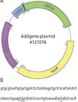
- Purified plasmids containing His-tagged biosensors for lactose/IPTG inducible expression (e.g., the calcium sensor MatryoshCaMP6 (Ast et al., 2017) in the vector pRSET-B) for nickel-nitrilotriacetic acid (Ni-NTA) affinity chromatography, as well as an empty vector, or the biosensor backbone protein lacking FPs (negative control) for expression in BL21 GOLD (DE3) cells. Plasmids and strains are described in more detail below
- Strains
Escherichia coli BL21 GOLD (DE3) [fhuA2 [lon] ompT gal (λ DE3) [dcm] ∆hsdS; λ DE3 = λ sBamHIo ∆EcoRI-B int::(lacI::PlacUV5::T7 gene1) i21 ∆nin5] (New England Biolabs, catalog number: C2527I ) - cOmpleteTM ULTRA Tablets, Mini, EDTA-free protease inhibitor cocktail (Merck, catalog number: 11836170001 )
- Ni-NTA Agarose Beads (Qiagen, catalog number: 30210 )
- Bradford assay kit (Bio-Rad, catalog number: 500-0006 )
- MOPS (3-(N-morpholino)propanesulfonic acid) buffer (Carl Roth, catalog number: 6979.3 )
- Imidazole (AppliChem, catalog number: A1073 ,0500; UN3263)
- D-(+) glucose monohydrate (Sigma-Aldrich, catalog number: 14431-43-7 )
- Lactose (Merk, catalog number: 7660.025 0)
- BSA (Bovine Serum Albumin; Carl Roth, catalog number: 0 163.3 )
- BactoTM Tryptone (BD BioSciences, catalog number: 211705 )
- BactoTM Yeast Extract (BD BioSciences, catalog number: 212750 )
- Sodium chloride (Carl Roth, catalog number: 3957.1 )
- BactoTM Agar (BD Biosciences, catalog number: 214030 )
- Antibiotic(s) required for plasmid selection. For example: ampicillin for vector pRSET-B containing MatryoshCaMP6 (Sigma-Aldrich, catalog number: 69-53-4 )
- Sodium hydroxide (Sigma-Aldrich, catalog number: 1310-73-2 )
- Calcium chloride (Sigma-Aldrich, catalog number: 10043-52-4 )
- 4-20% Mini-PROTEAN® TGXTM Precast Protein Gels, 10-well, 50 µl (Bio-Rad, catalog number: 4561094 )
- 4x SDS Sample buffer (MERK, catalog number: 70607-3 )
- PageRuler Prestained Protein Ladder (Thermo Fisher Scientific, catalog number: 26616 )
- PierceTM 6xHis Protein Tag Stain Reagent Set (Thermo Fisher Scientific, catalog number: 24570 )
- Lysogeny broth (LB) liquid medium with antibiotics (see Recipes)
- LB solid medium (see Recipes)
- Auto-induction medium (for composition and explanations, see Recipes)
- Lysis buffer (see Recipes)
- Wash buffer (see Recipes)
- Elution buffer (see Recipes)
- Final buffer (see Recipes)
- Ligand binding assay buffer (see Recipes)
- Coomassie stain buffer (see Recipes)
- Stock solution of carbenicillin (see Recipes)
- Running Buffer (see Recipes)
Equipment
- Standard micropipette or multichannel (8 or 12 channel) pipette (for 100 µl) (e.g., Sartorius AG, catalog number: 725240 )
- pH meter (InoLab® pH Level1, catalog number: 72.690.001 )
- Sonicator (Branson Sonifier cell disruptor B15)
- Centrifuge (Hettich Rotanta 460/460R )
- Microplate reader with adjustable bandwidths (Spark®, Tecan)
- Fluorescence stereomicroscope for large fields observations (ZEISS, model: Axio Zoom.V16 )
- Eppendorf ThermoMixer® C (Eppendorf, catalog number: 5382000015 )
- Autoclave (Systec GmbH, model: Systec V-150 )
- Mini-PROTEAN® Tetra Vertical Electrophoresis Cell (Bio-Rad, catalog number: 1658004 )
- NanoDrop 2000 (LabX, catalog number: LV40609601 )
- Eppendorf ThermoMixer® C (Eppendorf, catalog number: 5382000015 )
- Eppendorf Thermoblock SmartBlockTM 1.5 ml (Eppendorf, catalog number: 5360000038 )
- Gel Doc XR+ Gel Documentation System (Bio-Rad, catalog number: 1708195 )
Software
- ecan-related software (Spark®, Tecan Life Sciences)
- Excel (Microsoft, Microsoft Office Professional 2016)
- MyCurveFit 2019 (MyCurveFit, MyAssays Limited)
Procedure
文章信息
版权信息
© 2020 The Authors; exclusive licensee Bio-protocol LLC.
如何引用
Sadoine, M., Castro-Rodríguez, V., Poloczek, T., Javot, H., Sunal, E., Wudick, M. M. and Frommer, W. B. (2020). Affinity Purification of GO-Matryoshka Biosensors from E. coli for Quantitative Ratiometric Fluorescence Analyses. Bio-protocol 10(19): e3773. DOI: 10.21769/BioProtoc.3773.
分类
细胞生物学 > 基于细胞的分析方法 > 蛋白互作
分子生物学 > 蛋白质 > 表达
您对这篇实验方法有问题吗?
在此处发布您的问题,我们将邀请本文作者来回答。同时,我们会将您的问题发布到Bio-protocol Exchange,以便寻求社区成员的帮助。