

Method for Prolonged Incubation of Brain Slices
脑片长时间孵育方法的研究


发布: 2020年07月20日第10卷第14期 DOI: 10.21769/BioProtoc.3683 浏览次数: 5239
评审: Ehsan KheradpezhouhAnonymous reviewer(s)
参见作者原研究论文
The authors used this protocol in:

May 2016
Advertisement


Abstract
Slices of neuronal tissue maintain a high degree of topographical and functional properties of neurons and glia and therefore are extensively used for measurements of neuronal activity at the molecular, cellular and network levels. However, the lifespan of slice preparations is narrow, averaging of 6-8 hours. Moreover, the average viability of brain slices varies according to animal age and region of interest, leading to the high variability and low reproducibility of recorded data.
Previous techniques to increase the viability of brain slices focused on reducing cytotoxicity by chemical means, including alterations of the artificial cerebrospinal fluid (aCSF) composition to alleviate the direct damage of the slicing procedure or adding protective antioxidants to reduce cellular deterioration. In this protocol, we use a combination of hypothermia with firm control of the aCSF conditions in the recovery chamber (pH, temperature, and bacteria levels) to extend the slice viability significantly.
Given the breadth of its usage, improving slice viability and longevity can considerably increase data reproducibility and reduce the cost, time, and number of animals used in neurophysiological studies.
Background
Neuronal tissue slices provide unique advantages in basic neuroscience research as it allows direct investigation of neuronal network activity and the influence of pharmacological compounds on the central nervous system. Hence, it is imperative to prepare and maintain slices viability at the highest quality for experimental purposes. The viability of neuronal slices relies on several intrinsic and extrinsic factors (Buskila et al., 2014), which can be divided into three periods according to the slicing procedure: before (pre-slicing), during (slicing) and after (post-slicing) slicing procedure. The main factors that decrease slice viability in the pre-slicing period include ischemia and hypoxia occurring due to elimination of continuous blood supply (Buskila et al., 2020). Both hypoxia and ischemia, starting seconds after decapitation, lead to excitotoxic processes such as mitochondrial dysfunction and Ca2+ influx, which contribute to neuronal hyperexcitability and decrease in slice viability. The slicing procedure itself leads to physical damage of the semi-permeable membrane of neurons, resulting in neurotransmitter spill-over and inflow of compounds (i.e., Ca2+, Na2+, etc.) into the cell, which enhances neuronal cytotoxicity (Reid et al., 1988). The main factors affecting the slice viability during the post-slicing episode includes the temperature of the incubation solution and the stability of its pH, as these factors affect processes which impact the cells deterioration rate, including redox metabolism and the reduction in the oxygen transfer into the deep layers of the tissue (Reid et al., 1988). For long-term incubation of the slices (> 4 h), the level of bacteria is also an essential factor, as it contributes to slice deterioration via releasing of endotoxins, glial activation and neurodegeneration (Qin et al., 2007; Breen and Buskila, 2014).
To reduce the impact of the factors affecting the viability of the slices, we capitalize on previous protocols that found effective in increasing slice viability by reducing excitotoxicity during the pre-slicing and slicing steps and additionally use an active recovery (post-slicing) incubation chamber, the BraincubatorTM, which controls several parameters, such as temperature, pH and bacteria levels. In this system, slices are kept in a temperature-controlled chamber, while the aCSF flows through a secondary tank exposed to an ultraviolet C (UVC) light source that eradicates bacteria. The design and use of the BraincubatorTM have been described previously (Breen and Buskila, 2014; Buskila et al., 2014; Cameron et al., 2016 and 2017). Hypothermia has been proven highly efficient in reducing the metabolic rate and post-slicing cellular damage, hence, we have incubated brain slices at 15-16 °C. The combination of hypothermia with the UVC filtration system can extend slice viability for > 24 h (Buskila et al., 2014).
Materials and Reagents
- Masking tape (3M; RS components, catalog number: 124-0913 )
- Butterfly needles (27G; Global Medical Supply, catalog number: 91508T )
- Isoflurane (VCA I.S.O. 250 ml)
- Ketamine/Xylazine (100 mg/kg/10 mg/kg)
- Gel superglue (Loctite, 3 g)
- Carbogen (5% CO2 mixed in 95% O2; BOC Australia)
- N-methyl D-glucamine (NMDG; Sigma, catalog number: M2004-500G ), stored at RT
- KCl (Sigma, catalog number: P9333-1KG ), stored at RT
- NaH2PO4 monobasic monohydrate (Sigma, catalog number: S9638-25G ), stored at RT
- NaHCO3 (Sigma, catalog number: S5761-1KG ), stored at RT
- D-glucose (Sigma, catalog number: G5767-500G ), stored at RT
- MgSO4 (Sigma, catalog number: M7506-500G ), stored at RT
- CaCl2 (Sigma, catalog number: 223506-500G ), stored at RT
- HEPES (Sigma, catalog number: H3375-250G ), stored at RT
- Thiourea (Sigma, catalog number: T7875-100G ), stored at 4 °C
- Sodium L-ascorbate (Sigma, catalog number: 4034-100G ), stored at 4 °C
- Sodium pyruvate (Sigma, catalog number: P2256-25G ), stored at 4 °C
- NaCl (Sigma, catalog number: S5886-1KG ), stored at RT
- MgCl2 (Sigma, catalog number: M8266-100G ), stored at RT
- Perfusion aCSF (see Recipes)
- Incubation aCSF (see Recipes)
Equipment
- Anesthetic induction chamber (Darwall, catalog number: 1859 )
- 30 x 30 cm of polystyrene block (custom made)
- Large metal scissors (Fine Science Tools, catalog number: 14001-18 )
- Scalpel (Fine Science Tools, catalog number: 10004-13 )
- Scalpel blade (Fine Science Tools, catalog number: 10022-00 )
- Small scissors (Fine Science Tools, catalog number: 14088-10 )
- Forceps (Fine Science Tools, catalog number: 11271-30 )
- Spatula (Fine Science Tools, catalog number: 10090-13 )
- Hemostat (Fine Science Tools, catalog number: 13007-12 )
- Beaker (250 ml; Labdirect, catalog number: 355.018.250 )
- Vibratome (Leica Biosystems, model: VT1200S )
- Soft artist paint brush (size 2; Bunnings, catalog number: 1660207 )
- Pasteur pipette (Labdirect, catalog number: 88336 )
- BraincubatorTM (PaYo Scientific Ltd Pty; http://braincubator.com.au)
- Peristaltic pump (Masterflex, catalog number: EW-77122-20 )
- Tubing for Masterflex pump (Coleparmer, catalog number: EW-06508-15 )
- Small rodent guillotine (WPI)
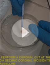
Procedure
文章信息
版权信息
© 2020 The Authors; exclusive licensee Bio-protocol LLC.
如何引用
Kekesi, O. and Buskila, Y. (2020). Method for Prolonged Incubation of Brain Slices. Bio-protocol 10(14): e3683. DOI: 10.21769/BioProtoc.3683.
分类
神经科学 > 基础技术 > 快速制片
神经科学 > 基础技术 > 低温贮藏
细胞生物学 > 组织分析 > 组织培养
您对这篇实验方法有问题吗?
在此处发布您的问题,我们将邀请本文作者来回答。同时,我们会将您的问题发布到Bio-protocol Exchange,以便寻求社区成员的帮助。