

Tyramide Signal-Amplified Immunofluorescence of MYCN and MYC in Human Tissue Specimens and Cell Line Cultures
酪胺信号放大的人体组织标本和细胞系培养物中的MYCN和MYC免疫荧光
发布: 2020年07月05日第10卷第13期 DOI: 10.21769/BioProtoc.3677 浏览次数: 4326
评审: Alak Mannasujan kumar mondalAnonymous reviewer(s)
参见作者原研究论文
The authors used this protocol in:

Mar 2020
Advertisement


Abstract
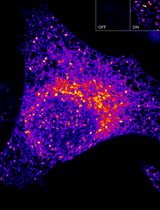
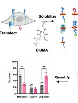
MYC family members, MYC, MYCN, and MYCL, are oncogenic transcription factors that regulate the expression of genes involved in normal development, cell growth, proliferation, metabolism, and survival. While MYC is amplified and/or overexpressed across a variety of tissue types, MYCN is often overexpressed in tumors of the nervous system (neuroblastoma and medulloblastoma) or with neuroendocrine features (neuroendocrine prostate cancer). Given recent reports that MYCN expression is also deregulated in a variety of non-neuronal tissue types, we investigated whether MYCN was also deregulated in triple-negative breast cancer (TNBC). In contrast to previous individual immuno-fluorescence (IF) stains against higher expressing MYC family isoform protein, we developed an IF stain to simultaneously detect both MYCN- and MYC-expressing cells within the same tumor cell population. Our methodology allows for the detection of low level MYCN and MYC expression and can be multiplexed with additional protein probes. Herein, using tyramide signal amplification (TSA), we present two protocols for the IF detection of MYCN and MYC on formalin-fixed paraffin embedded (FFPE) tumor sections and in cell lines fixed in situ after growth as adherent cultures on chambered microscope slides.
Keywords: Cancer biology (肿瘤生物学)Background
Previous studies have demonstrated MYCN and MYC preferentially regulate the same set of core genes involved in metabolism and cell growth, and while the MYCN allele can functionally replace MYC in murine development (Malynn et al., 2000), MYCN and MYC have separate temporal regulation over organogenesis in early vertebrate development (Hurlin, 2013). MYCN expression is essential for initial establishment of stem and progenitor populations; over the course of organ system development, MYCN expression switches to low MYC expression to support stem and progenitor cell maintenance, and during cell lineage commitment and expansion, elevated MYC levels drive highly proliferative cells until they reach terminal differentiation (Hurlin, 2013).
A similar relationship has been observed in the development of TNBC metastases where MYCN expression is elevated in newly seeded metastatic lesions that expand and differentiate into highly proliferative MYC-expressing tumors (Lawson et al., 2015). Given the near ubiquitous nature of MYC deregulation across tumor tissue types, MYC is the most studied MYC family member. However, due to a correlation between deregulated MYCN expression and a poor prognosis (Beltran, 2014), MYCN has been increasingly studied in both neuronal and non-neuronal cancers, including acute myeloid leukemia (Kawagoe et al., 2007), small-cell lung cancer (Funa et al., 1987), ovarian cancer (Baratta et al., 2015), and now TNBC (Schafer et al., 2020). Therefore, we began to explore the relationship between MYCN and MYC by developing an IF stain that could simultaneously detect both isoforms within the same tumor tissue section. Established protocols that involve the IF detection of MYCN or MYC have been separate stains that primarily recognizes each isoform at highly amplified/overexpressed levels. Given that TNBC and other non-neuronal tissue types exhibit aberrant low-level MYCN expression, we developed a method to detect lower expression of MYCN and MYC using TSA. Further, we have two protocol versions of the method: application of the dual MYC family isoform (MYCN and MYC) TSA-IF to FFPE tumor sections, and a modified version of the stain for evaluation MYC family isoforms expression in cell line cultures fixed in situ after growth as adherent cultures on chambered microscope slides. We anticipate both MYCN and MYC are coexpressed in a variety of cancer tissue types and the advent of these IF methodologies will allow investigators to further characterize MYCN- versus MYC-expressing cells in tumor development and disease etiology.
Materials and Reagents
- Microscopy slides (VWR, catalog number: 48311-703 )
- Coverslips (Fisherbrand, catalog number: 12-545K )
- HistoPrep pen (Fischer Scientific, catalog number: 14-905-30 )
- PAP pen (Electron Microscopy Sciences, catalog number: 71310 )
- 10% neutral buffered formalin (Thermo scientific, catalog number: 5701 ), store at room temperature
- Xylene (Fisher Scientific, catalog number: X3P-1GAL ), store at room temperature
- Ethanol (Pharmco, catalog number: 111000200 ), store at room temperature
- Citra plus antigen retrieval buffer (BioGenex, catalog number: HK080-9K ), store at 4 °C
- Phosphate-buffered saline (Gelifesciences, HyClone, catalog number: SH30013.03 ), store at 4 °C
- Tween-20 (Acros Organics, catalog number: 23336-0010 ), store at room temperature
- Triton X-100 (Millipore, OmniPur, catalog number: 9410 ), store at room temperature
- 30% hydrogen peroxide (H2O2) (Fisher Chemical, catalog number: H325-100 ), store at 4 °C
- Image-iT FX Signal Enhancer (Invitrogen, catalog number: I36933 ), store at 4 °C
- Goat serum (Gemini, catalog number: 100-109 ), store at -20 °C
- DMSO (Sigma, catalog number: D2650-100ml ), store at room temperature
- FITC TSA reagent (PerkinElmer, catalog number: NEL741B001KT ), store at 4 °C
- Cy3 TSA reagent (PerkinElmer, catalog number: NEL744B001KT ), store at 4 °C
- Optional: Cy5 TSA reagent (PerkinElmer, catalog number: NEL745B001KT ), store at 4 °C
- DAPI (Sigma-Aldrich, catalog number: D9542 ), store at -20 °C, or Hoechst (Invitrogen, catalog number: H3570 ), store at 4 °C
- SlowFade Gold Antifade Mountant (ThermoFisher Scientific, catalog number: S36937 ), store at room temperature
- Nail polish (any kind or brand will suffice)
- Antibodies
- Permeabilization buffer (see Recipes)
- Blocking buffer (see Recipes)
- Antibody diluent (see Recipes)
- Wash buffer (see Recipes)
- FITC signal amplification buffer (see Recipes)
- Cy3 signal amplification buffer (see Recipes)
- Eight-well chamber microscope slides (Corning, catalog number: 354108 )
- Methanol (Millipore, catalog number: MX0485-7 ), store at room temperature
- Antibodies
Equipment
- Slide Warmer (e.g., Lab-Line Instruments, model: CPC-600N1 )
- Electric pressure cooker (e.g., Cuisinart, model: CPC-600N1 )
- Microscope slide moisture chamber (e.g., Ted Pella, catalog number: 21053 )
- Slide staining station (e.g., Tissue-Tek, catalog number: 4451 )
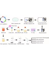
Procedure
文章信息
版权信息
© 2020 The Authors; exclusive licensee Bio-protocol LLC.
如何引用
Readers should cite both the Bio-protocol article and the original research article where this protocol was used:
- Schafer, J. M. and Pietenpol, J. A. (2020). Tyramide Signal-Amplified Immunofluorescence of MYCN and MYC in Human Tissue Specimens and Cell Line Cultures. Bio-protocol 10(13): e3677. DOI: 10.21769/BioProtoc.3677.
- Schafer, J. S., Lehmann, B. D., Gonzalez-Ericsson, P. I., Marshall, C. B., Beeler, J. S., Redman, L. N., Jin, H., Sanchez, V., Stubbs, M. C., Scherle, P., Johnson, K. N., Sheng, Q., Roland, J. T., Bauer, J. A., Shyr, Y., Chakravarthy, B., Mobley, B. C., Hiebert, S. W., Balko, J. M., Sanders, M. E., Liu, P. C. C., Pietenpol, J. A. (2020). Targeting MYCN-expressing triple-negative breast cancer with BET and MEK inhibitors. Sci Transl Med 12(534).
分类
癌症生物学 > 癌症生物化学 > 蛋白质
生物化学 > 蛋白质 > 荧光
您对这篇实验方法有问题吗?
在此处发布您的问题,我们将邀请本文作者来回答。同时,我们会将您的问题发布到Bio-protocol Exchange,以便寻求社区成员的帮助。