

Organotypic Slice Culture of the Embryonic Mouse Brain
胚胎小鼠大脑的器官型切片培养
发布: 2020年07月05日第10卷第13期 DOI: 10.21769/BioProtoc.3674 浏览次数: 5916
评审: Geoffrey C. Y. LauWoojong LeeLinlin Sun
参见作者原研究论文
The authors used this protocol in:
Feb 2019
Advertisement


Abstract
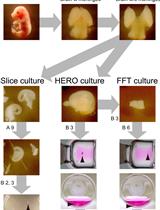
Organotypic slice culture is a powerful technique for exploring the embryonic development of the mammalian brain. In this protocol we describe a basic slice culture technique we have used for two sets of experiments: axon guidance transplant assays and bead culture assays.
Keywords: Organotypic (器官型)Background
Organotypic slice culture is a technique that has been widely used in recent years and has been a particularly popular technique in the field of neurodevelopment. It has the great advantage of allowing the culture of developing neural cells in-vitro while maintaining the in-vivo structure of the tissue. In this protocol we describe the slice culture technique we used for two experiments from our recent paper (Clegg et al., 2019). Firstly we performed an axon guidance transplant assay whereby fluorescently labeled tissue was transplanted into a non-fluorescent host slice in order to observe axon outgrowth. This is a modified version of a protocol previously used to study the development of the corpus callosum, but could be easily adapted to examine other axon tracts such as the thalamocortical tract (Niquille et al., 2009). Secondly, we performed a bead culture assay in which beads soaked in recombinant FGF protein was embedded in the tissue, this allowed the examination of the molecular and cellular response of the tissue to the FGF protein. The use of protein soaked beads in this way provides a method for focal delivery of recombinant protein at a specific position, in contrast to bath application which exposes all tissue to the protein equally. This mimics the situation in-vivo where a morphogen (such as FGF17) is expressed at a particular anatomical position and then diffuses through the tissue. This technique could be used to explore the response to a number of different recombinant proteins or pharmacological agents.
Materials and Reagents
Materials
- 7 ml bijou tubes (Greiner, catalog number: 189170 )
- Pipette tips (Greiner, catalog number: 739288 )
- 1.5 ml microcentrifuge tubes (Greiner, catalog number: 616201 )
- Peel-A-Way® Embedding Molds (Square–S22, Polysciences)
- Falcon® 60 mm TC-treated Center Well Organ Culture Dish (Corning, catalog number: 353037 )
- 13 mm Nuclepore polycarbonate track-etched membranes (Whatman, catalog number: 110401 )
- Specimen disk
- Adhesive
Animals
Mice used in our original study ubiquitously express τGFP and have been described previously (Pratt et al., 2000). All mice were maintained on a CD-1 background (Charles River, strain code: 022). Mice used for timed matings were aged between 6 and 24 weeks.
Reagents
- Earls balanced salt solution (EBSS) calcium, magnesium, phenol red (Thermo Fisher, catalog number: 24010043 )
- SeaPlaque GTG Agarose (Lonza, catalog number: 50110 )
- Affi-Gel Blue gel beads (Bio-Rad, catalog number: 1537301 )
- MEM (Thermo Fisher, catalog number: 11090081 )
- Neurobasal medium (Thermo Fisher, catalog number: 21103049 )
- 1 M HEPES (Thermo Fisher, catalog number: 15630080 )
- Penicillin-streptomycin (Thermo Fisher, catalog number: 15140122 )
- D-Glucose solution (Merck, catalog number: G8769 )
- Gentamicin 50 mg/ml (Thermo Fisher, catalog number: 15750060 )
- Glutamine 200 mM (Thermo Fisher, catalog number: 25030081 )
- Fetal bovine Serum (Merck, catalog number: F4135 )
- B-27 supplement (Thermo Fisher, catalog number: 17504044 )
- OCT embedding matrix (Cellpath, catalog number: KMA-0100-00A )
- 1x Krebs buffer (from 10x stock, see recipes)
- 1x PBS (Thermo Fisher catalog number: 14190094 )
- NaCl (Thermo Fisher catalog number: 10428420 )
- KCl (Thermo Fisher catalog number: 10684732 )
- NaH2PO4 (Thermo Fisher catalog number: 10723621 )
- CaCl2 (Thermo Fisher catalog number: 10657662 )
- MgCl2 (Thermo Fisher catalog number: 10386743 )
- Triton X-100 (Merck, catalog number: 10789704001 )
- Bovine serum albumin (Merck, catalog number: A1933 )
- Anti-GFP antibody (Abcam, catalog number: ab290 )
- Goat anti-Rabbit IgG 488 (Thermo Fisher, catalog number: A-11008 )
- DAPI (Thermo Fisher, catalog number: D1306 )
- Vectashield Hardset (VectorLabs, catalog number: H-1400 )
- 10x Krebs Buffer (see Recipes)
- 1x Krebs with antibiotic (see Recipes)
- MEM with serum (see Recipes)
- Neurobasal (see Recipes)
Equipment
- Pipettes
- Laminar flow tissue culture hood
- Tissue culture incubator (37 °C, 5% CO2)
- Dissecting microscope (Euromex, catalog number: DZ1100 )
- Leica VT1000 S vibratome
- Water bath
- Fisherbrand microspatula with flat-ended blade (Thermo Fisher, catalog number: 21-401-20 )
- Forceps, size 5 (Fine Science Tools, catalog number: 11251-20 )
- Microscissors, 5 mm cutting edge (Fine Science Tools, catalog number: 15003-08 )
- Scissors, 9 cm (Fine Science Tools, catalog number: 14060-09 )
- 45° Sterile blade (Altomed, model: A10136 )
- Cryostat (Leica, catalog number: CM3050 S )
- Super-frost Plus slides (Thermo Fisher, catalog number: J1800AMNZ )
- Hydrophobic barrier pen (VectorLabs, catalog number: H-4000 )
Procedure
文章信息
版权信息
© 2020 The Authors; exclusive licensee Bio-protocol LLC.
如何引用
Readers should cite both the Bio-protocol article and the original research article where this protocol was used:
- Clegg, J. M. and Pratt, T. (2020). Organotypic Slice Culture of the Embryonic Mouse Brain. Bio-protocol 10(13): e3674. DOI: 10.21769/BioProtoc.3674.
- Clegg, J. M., Parkin, H. M., Mason, J. O. and Pratt, T. (2019). Heparan sulfate sulfation by Hs2st restricts astroglial precursor somal translocation in developing mouse forebrain by a non-cell-autonomous mechanism. J Neurosci 39(8): 1386-1404.
分类
神经科学 > 发育 > 外植体培养
发育生物学 > 细胞信号传导 > 配体
细胞生物学 > 细胞信号传导 > 发育
您对这篇实验方法有问题吗?
在此处发布您的问题,我们将邀请本文作者来回答。同时,我们会将您的问题发布到Bio-protocol Exchange,以便寻求社区成员的帮助。