

Preparation of Drosophila Polytene Chromosomes, Followed by Immunofluorescence Analysis of Chromatin Structure by Multi-fluorescence Correlations
制备果蝇多线染色体并通过多重荧光关联对染色质结构进行免疫荧光分析
发布: 2020年07月05日第10卷第13期 DOI: 10.21769/BioProtoc.3673 浏览次数: 9989
评审: Imre GáspárRumen IvanovAnonymous reviewer(s)
参见作者原研究论文
The authors used this protocol in:

Sep 2019
Advertisement


Abstract
Drosophila larval salivary gland polytene chromosome squashes have been used for decades to analyze genome-wide protein-binding patterns, transcriptional activation processes, and changes in chromatin structure at specific genetic loci. There have been many evolutions of the squashing protocol over the years, with sub-optimal reproducibility and low sample success rate as accepted caveats. However, low sample success rates are an obvious disadvantage when polytene chromosomes are used for more high-throughput approaches, such as genetic or antibody screens, or for experiments requiring high-quality chromosome structure preservation. Here we present an exceptionally reproducible squashing and fluorescence staining protocol, which generates high-quality fluorescence images on well-spread chromosomes. This is followed by our novel, semi-automated MATLAB analysis program used to determine correlations between fluorescence signals of interest at a single site on polytene chromosomes, in a pixel-by-pixel manner. In our case, we have used this approach to assess chromatin changes at genomic sites, ectopically targeted by nuclear pore proteins. The use of our analysis program increases the ability to make unbiased conclusions on changes in chromatin structure, or in protein recruitment to chromatin, regardless of sample variation in immunofluorescence staining. As it is simply based upon differences in fluorescence intensity at a defined location, the provided analysis program is not limited to analysis of polytene chromosome, and could be applied to many different contexts where correlation between fluorescent signals at any particular location is of interest.
Keywords: Polytene chromosome squashes (多线染色体按压)Background
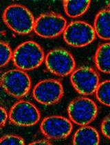
Polytene chromosomes from Drosophila larval salivary glands have an unusually large size (visible under a typical cell-culture microscope). This is due to altered cell cycle processes that result in ~1,000 copies of each chromosome that do not segregate from each other. The resulting large chromosomes have stereotyped and reproducible alternating bands of condensed and decondensed chromatin, as chromosomes are aligned and locus-specific chromatin structure is visible upon polytenization. The nature of these chromosomes makes the analysis of chromatin structure uniquely visible relative to diploid cell types, enabling analysis of chromatin changes throughout development, transcriptional activation, or upon perturbation. Additionally, genome-wide and site-specific chromatin binding of proteins can be visualized by immunofluorescence staining of polytene chromosomes (Paro, 2008; Cai et al., 2010), allowing for investigation of the relationships between candidate proteins and chromatin condensation, transcriptional activity, and histone modifications. In this manner, polytene chromosome staining provides a rapid and inexpensive alternative or supplement to molecular assays such as Chromatin Immunoprecipitation, coupled to sequencing (ChIP-seq).
Our squashing and fluorescence analysis pipeline was specifically designed for analysis of chromatin structure and protein recruitment/changes at single bands of interest, originally for a band of LacI-tethered protein bound to a genomic lacO repeat array (Kuhn et al., 2019). In this study, we targeted specific nuclear pore components, fused to LacI, to lacO arrays, integrated at defined sites in the genome. We then aimed to determine whether such targeting leads to recruitment of candidate interacting partners, and also to changes in target chromatin compaction. This approach involved identification of a single band of interest (harboring the lacO site) within an entire genome of spread chromosomes, which required high-quality chromosome spreading for increased visibility and analysis of chromosome banding patterns. Additionally, screening through multiple antibodies to assay for many candidate interacting partners required an extremely reproducible squashing protocol, with minimal sample failure rate. To achieve these goals, we developed this optimized polytene squashing protocol, which can be utilized to improve image quality and sample success rate. It is also especially useful for chromosome ‘mappability’ (determining genomic location by cytological mapping of polytene banding patterns) and for more high-throughput applications.
In order to reliably analyze site-specific changes in chromatin structure (i.e., changes in chromatin condensation as assessed by DNA stain fluorescence intensity), and in protein recruitment, we developed a semi-automated MATLAB function. This function, PCCcalc, calculates the Pearson Correlation Coefficients (PCC) between multiple fluorescence signals at a defined genomic site on a pixel-by-pixel basis in an unbiased manner. This greatly reduces data analysis time and subjectivity derived from manual analysis and user-based variability. Additionally, this approach circumvents the impact of slide-to-slide variability in fluorescence background or image acquisition, as the analysis focuses on the relationship between fluorescence signals more than absolute values of a given signal. This method can be used on any easily identifiable site on polytene chromosomes to determine binding correlations between multiple protein factors, or to find correlations between protein of interests and associated changes in chromatin compaction. Furthermore, we believe this analysis method can be extended to determine fluorescence correlation beyond the described use in polytene chromosomes, and can be used to analyze co-localization or co-occurrence of fluorescently labeled biological markers in many other contexts and cell types.
Materials and Reagents
- Filter paper (Fisherbrand, catalog number: 05-714-4 )
- Hammer with a rubber head (Graham Field, catalog number: 1312-1 )
- Fine sharpie
- 1.5 ml tubes (USA Scientific, catalog number: 1615-5510 )
- Dissection Well Dish/Pyrex Spot Plate (Fisher Scientific, catalog number: 13-748B )
- Dumont Fine #55 Forceps (Fine Sceince Tools, catalog number: 11295-51 )
- Coverslips (Fisherbrand, catalog number: 12-542A )
- Coplin Jar (VWR, catalog number: 74830-150 )
- Humidity chamber (such as Pyrex Lidded Glass Baking Dish)
- Poly-L-Lysine-treated Slides (Polysciences, catalog number: 22247 )
- Poly-L-Lysine (alternative to pre-treated slides above) (Sigma-Aldrich, catalog number: P8920-100 ml )
- Sigmacote (Sigma-Aldrich, catalog number: SL2-100ML )
- Phosphate Buffered Saline (PBS) reagents:
- NaCl (Fisher, catalog number: BP358-212 )
- KCl (Fisher, catalog number: BP366-500 )
- Na2HPO4 (Sigma, catalog number: S3264-500G )
- KH2PO4 (Sigma, catalog number: P9791-1KG )
- Primary Antibodies used: anti-LacI (Rockland, catalog number: 600-401-B05 ), anti-Nup98 (Capelson et al., 2010), anti-Brm (Nakayama et al., 2012)
- Tween 20 (Fisher, catalog number: BP337-500 )
- Glacial Acetic Acid (Fisher, catalog number: BP2401-212 )
- 16% Paraformaldehyde (PFA) (Alfa Aesar, catalog number: 43368 )
- Liquid nitrogen
- Kimwipes or other tissue-style wiper (Fisher Scientific, catalog number: 06-666A )
- Fluorescent Secondary Antibodies (e.g., Thermo Fisher, catalog number: A-11004 )
- Hoechst 33342 (Thermo Fisher, catalog number: H3570 ) (10 mg/ml stock)
- Prolong Gold Antifade (Thermo Fisher, catalog number: P36930 )
- PBS (see Recipes)
- PBST (see Recipes)
- Fixation solution (see Recipes)
- 45% acetic acid (see Recipes)
- Blocking solution/Antibody cocktail base (see Recipes)
Equipment
- Forceps
- Pipettes
- Chemical hood
- Dissecting Microscope (such as Leica, model: Leica S6E )
- Cell Culture/Phase Contrast Microscope (such as Leica, model: Leica DM IL )
- Widefield epifluorescent microscope (such as Leica, model: Leica DM6000 ) and/or laser scanning confocal microscope
- (Optional) Orbital shaker (VWR, catalog number:12620-938)
Software
- MATLAB (license) (necessary for analysis)
- Graphing/Statistical Analysis software, e.g., Excel, Prism (licenses) (necessary for analysis)
- (Optional) ImageJ (free)
Procedure
文章信息
版权信息
© 2020 The Authors; exclusive licensee Bio-protocol LLC.
如何引用
Readers should cite both the Bio-protocol article and the original research article where this protocol was used:
- Kuhn, T. M., Little, S. C. and Capelson, M. (2020). Preparation of Drosophila Polytene Chromosomes, Followed by Immunofluorescence Analysis of Chromatin Structure by Multi-fluorescence Correlations. Bio-protocol 10(13): e3673. DOI: 10.21769/BioProtoc.3673.
- Kuhn, T. M., Pascual-Garcia, P., Gozalo, A., Little, S. C. and Capelson, M. (2019). Chromatin targeting of nuclear pore proteins induces chromatin decondensation. J Cell Biol 218(9): 2945-2961.
分类
生物化学 > 蛋白质 > 荧光
细胞生物学 > 细胞成像 > 荧光
您对这篇实验方法有问题吗?
在此处发布您的问题,我们将邀请本文作者来回答。同时,我们会将您的问题发布到Bio-protocol Exchange,以便寻求社区成员的帮助。