

Single Cell Migration Assay Using Human Breast Cancer MDA-MB-231 Cell Line
利用人乳腺癌MDA-MB-231细胞系进行单细胞迁移分析

发布: 2020年04月20日第10卷第8期 DOI: 10.21769/BioProtoc.3586 浏览次数: 7518
评审: Gal HaimovichChaitali BasolePhilipp Wörsdörfer
参见作者原研究论文
The authors used this protocol in:

Apr 2019
Advertisement


Abstract
Cell migration is a fundamental cellular process that plays a crucial role in many physioglogical and pathological processes such as wound healing or cancer metastasis. Many assays have been developed to examine cell migration, such as the wound healing or scratch assay, Boyden Chamber or transwell assay, and the method we will describe here, single cell migration assay. In this assay, cells are plated sparsely on a collagen coated plate and live cell imaging is performed over a period of 2 h at 1 frame per minute. After imaging is completed, cells are tracked manually using ImageJ by tracking movement of the centroid of the cell. These data points are then exported and overall distance travelled from frame to frame is determined and divided by total time imaged to determine speed of the cell. This method provides a quick way to examine effect of cellular manipulation on cell migration before proceeding to perform more complex assays.
Keywords: Single cell migration (单细胞迁移)Background
Cell migration plays an important role in both physiological and pathological processes ranging from embryonic development to angiogenesis and tumor metastasis (Le Clainche and Carlier, 2008). Cell motility is a highly orchestrated event that can be summarized as a cycle of four basic steps: i) membrane protrusion driven by actin polymerization, ii) stabilization of protrusion through integrin-mediated cell-matrix adhesion, iii) cell-body translocation driven by actomyosin contractile force, and finally, iv) rear release as a result of the mechanical action of contractile force and/or proteolysis of cell-matrix adhesion components (Sheetz et al., 1999; Ridley et al., 2003; Panetti et al., 2004; Stradal and Scita, 2006; Tomasevic et al., 2007; Le Clainche and Carlier, 2008). These processes involve dynamic remodeling of actin cytoskeleton which is dependent on de novo synthesis as well as regulation of important structural and regulatory components of actin cytoskeletal system.
Several methods have been developed to examine cell migration and can be separated into 2D and 3D assays. 2D migration assay have their advantages as they are typically easier and quicker to perform, however, lack the physiological representation that 3D assays may provide. We will provide a brief description of some of the other methods for cell migration. The scratch assay or wound healing assay is a commonly used technique to evaluate directed cell migration (Cory, 2011). In this assay, cells are plated to form a confluent monolayer and a stratch is introduced in the monolayer. The cell-free zone is then monitored as adjacent cells migrate to close the gap. While simple to perform, cell proliferation may bias the readout of this assay as that can influence scratch closure. In addition, cells in this assay are forced to move in one direction. The Boyden Chamber assay or transwell assay is another technique used to evaluate cell migration (Falasca et al., 2011). In this assay, cells are placed on one side of a porous membrane and allowed to migrate through the pores to the other side. An advantage of this assay is it allows for chemotaxis and works for both adherent and non-adherent cells. As with the scratch assay, migration in this assay is defined. Some disadvantages of this assay include difficulty to visualize cells and morphology due to transitive state of cells while migrating through pores.
In this protocol, we will describe the use of single cell migration assay to examine MDA-MB-231 cell migration. The key difference in this protocol is that we examine random cell migration rather than directed migration. This allows us to examine cell migration in a manner that is less influenced by cell-cell contact and its innate ability for directional persistence, i.e., ability to migrate randomly in a single direction. While obviously not a true physiological representation of in vivo cell migration, this assay allows us to quickly analyze effect of cellular manipulation on its ability to migrate. While this assay uses MDA-MB-231, this protocol can be performed with any migratory cell line. We have performed this assay using multiple cell lines with minor modifications (media used and amount of cells plated).
Materials and Reagents
- 24-well cell culture plate (Corning, Costar, catalog number: 9761146 )
- 100 mm cell culture plate (Corning, catalog number: 08-772-22 )
- 15 ml conical centrifuge tubes (Falcon, catalog number: 14-959-49D )
- 100% CO2 Tank
- DMEM (Lonza, BioWhittaker, catalog number: BW12-604F )
- Heat-inactivated FBS (Corning, catalog number: 35011CV )
- 100x Antibiotic-Antimycotic (Gibco, catalog number: 15240-062 )
Note: This is optional. We use this as extra assurance to prevent bacterial and fungal contamination in our cell culture. For some cell lines, this can hinder cell growth. - 100x Sodium pyruvate (Gibco, catalog number: 11360-070 )
- Collagen I, 3 mg/ml (Gibco, catalog number: A1048301 )
- Trypsin (Lonza, Trypsin-Versene, catalog number: BW17161E )
- PBS (Lonza, BioWhittaker, catalog number: BW17516F )
- MDA-MB-231 (ATCC, catalog number: CRM-HTB-26 )
- DMEM S+ media (see Recipes)
- Collagen (10 μg/ml) (see Recipes)
Equipment
- Hemocytometer
- Live-cell imaging environmental chamber (Tokai Hit live-cell environmental chamber)
- Centrifuge (unrefrigerated) for 15 ml tubes (Sorvall, Legend RT, SO-LEGRT)
- Cell incubator set to 37 °C and 5% CO2
- Waterbath or drybath set to 37 °C (Boekel Scientific, Small Water Bath, 290100)
- Microscope with automatic stage control (Olympus IX71 Inverted Phase with Prior Scientific motorized stage) (Figure 1)
- Camera for microscope (Hamamatsu)
- 10x objective (Olympus)
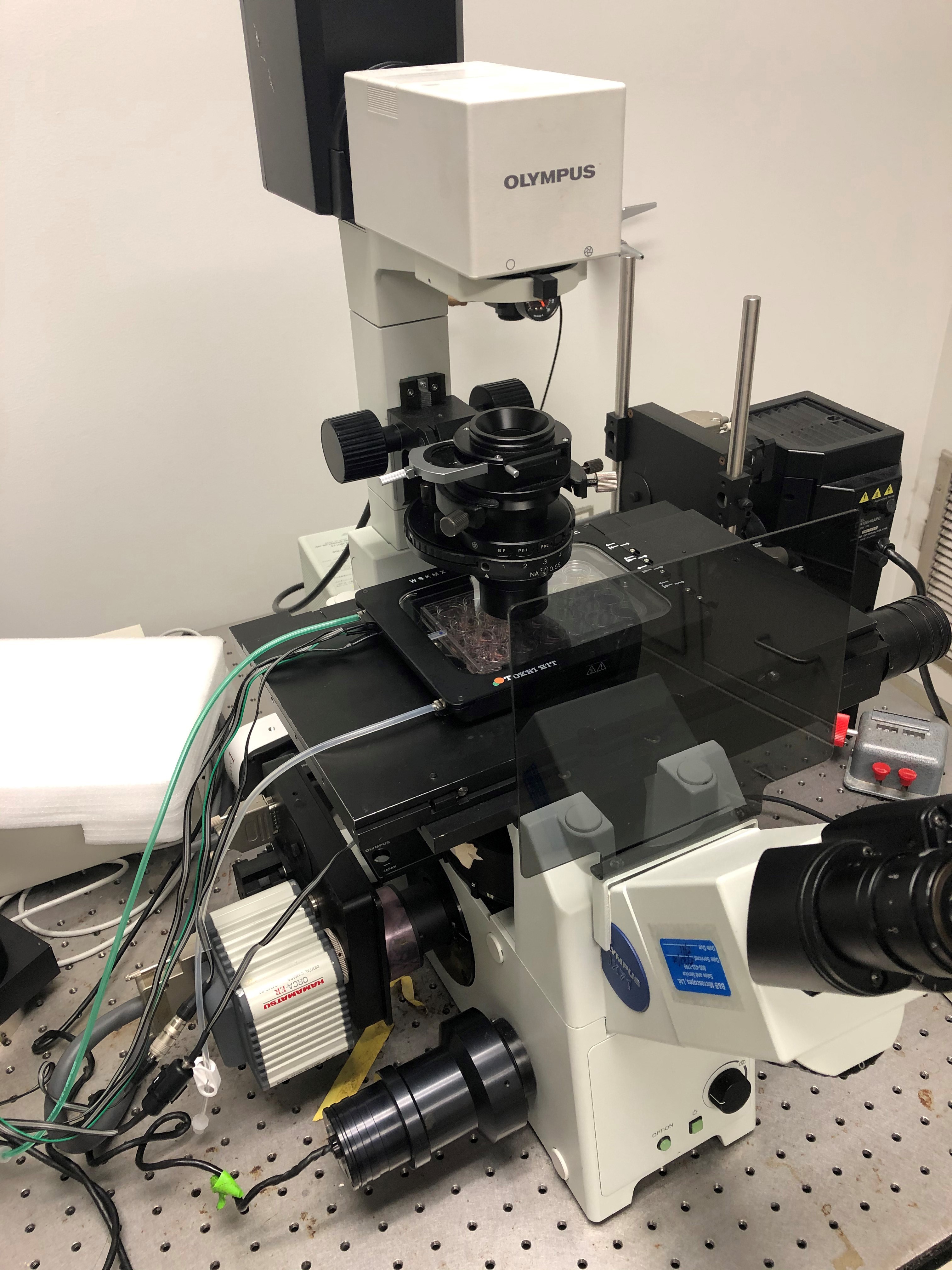
Figure 1. Example of a complete setup for cell tracking using Olympus IX71 inverted microscope, Prior mechanized stage, and Tokai Hit environmental chamber
Software
- Software compatible with microscope for time-lapse image aquisition (Olympus, cellSens)
- Fiji ImageJ (https://fiji.sc/)
- Microsoft Excel
Procedure
文章信息
版权信息
© 2020 The Authors; exclusive licensee Bio-protocol LLC.
如何引用
Readers should cite both the Bio-protocol article and the original research article where this protocol was used:
- Gau, D. M. and Roy, P. (2020). Single Cell Migration Assay Using Human Breast Cancer MDA-MB-231 Cell Line. Bio-protocol 10(8): e3586. DOI: 10.21769/BioProtoc.3586.
- Gau, D., Veon, W., Shroff, S. G. and Roy, P. (2019). The VASP-profilin1 (Pfn1) interaction is critical for efficient cell migration and is regulated by cell-substrate adhesion in a PKA-dependent manner. J Biol Chem 294(17): 6972-6985.
分类
癌症生物学 > 侵袭和转移 > 细胞生物学试验 > 细胞迁移
细胞生物学 > 细胞运动 > 细胞迁移
细胞生物学 > 细胞成像 > 活细胞成像
您对这篇实验方法有问题吗?
在此处发布您的问题,我们将邀请本文作者来回答。同时,我们会将您的问题发布到Bio-protocol Exchange,以便寻求社区成员的帮助。