- EN - English
- CN - 中文
Characterization of Immunological Niches within Peyer’s Patches by ex vivo Photoactivation and Flow Cytometry Analysis
体外光激活和流式细胞术分析派尔斑免疫龛位
发布: 2020年03月20日第10卷第6期 DOI: 10.21769/BioProtoc.3562 浏览次数: 5133
评审: mohan babuMeenal SinhaLuis Alberto Sánchez Vargas

相关实验方案

利用基于 FRET 的 SuperClomeleon 传感器监测器官型海马切片中细胞内氯离子水平变化
Sam de Kater [...] Corette J. Wierenga
2025年03月05日 2780 阅读
Abstract
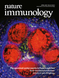
T follicular helper (Tfh) cells regulate B cell selection for entry into the germinal center (GC) reaction or for differentiation into antibody forming cells. This process takes place at the border between the T and B zones in lymphoid organs and involves physical contacts between T and B cells. During these interactions, T cells endow the B cells with selection signals that promote GC seeding or plasmablast differentiation based on their B cell receptor affinity. In Peyer’s patches (PPs), T cells promote B cell colonization of the subepithelial dome (SED) without effective affinity-based clonal selection. To specifically characterize the T cell population that resides within the SED niche, we performed ex vivo photoactivation of the SED compartment followed by flow cytometry analysis of the labeled cells, as described in this protocol. This technique integrates both spatial and cellular information in studies of immunological niches and can be adapted to various experimental systems.
Keywords: Photoactivation (光激活)Background
Affinity maturation, the process wherein the affinity of serum antibodies towards a specific antigen increases over time, is achieved by selection of B cells bearing high affinity BCRs within germinal centers (GCs). Increase in antibody affinity is mediated through iterative cycles of somatic hyper mutation and affinity-based selection, a process which is orchestrated by T follicular helper (Tfh) cells (Kepler and Perelson, 1993; Oprea and Perelson, 1997; Victora and Nussenzweig, 2012). The GC is comprised of two microanatomical sites; the dark zone, where B cells proliferate and acquire somatic hypermutations, and the light zone, where B cells interact with cognate antigen and T cells. Although intravital imaging techniques were able to define immune cell dynamics in GCs (Allen et al., 2007; Hauser et al., 2007; Schwickert et al., 2007), transition between the GC zones remained in question. This problem was later solved by the generation of mice expressing photoactivatable GFP (PA-GFP) (Victora et al., 2010).
PA-GFP is a GFP variant whose peak excitation wavelength shifts from 415 nm (inactive PA-GFP) to 495 nm (active PA-GFP) upon two-photon irradiation at 830 nm. The non-activated PA-GFP is fluorescent as well, and this property can be used to distinguish between the photoactivated area and the total cells (Patterson and Lippincott-Schwartz, 2002).
The use of mice expressing PA-GFP provided direct evidence for interzonal migration in the GC and for the definition of the major GC exit zone (Victora et al., 2010; Stoler-Barak et al., 2019). Combination of intravital two-photon laser scanning microscopy with in situ photoactivation, allowed the microanatomical labeling of distinct niches within the germinal center, and led to the discovery that T cell help controls the movement between the two GC zones (Victora et al., 2010).
Cell-surface markers are commonly used to define a cell population in a specific niche; however, distinctive markers are not always available, generating a gap between the cellular and spatial information in the studied tissue. Furthermore, unknown cell populations that reside within a specific niche are usually hard to detect and characterize by conventional techniques. To overcome these limitations, photoactivation-based approaches have been used for unbiased identification of tissue-resident immune cells with minimal a priori knowledge of unique cell-surface marker expression. Niche-specific landmarks are often introduced into mice prior to labeling by photoactivation to define the area of interest. For example, adoptive cell transfer of fluorescently labeled B cells mark the B cell area within a tissue and can guide the selection of the region of interest for photoactivation (Medaglia et al., 2017). In the study associated with this protocol and as described here, we specifically photoactivated the subepithelial dome niche within the Peyer’s patch (Biram et al., 2019). This protocol can be adapted to other niches and additional tissues of interest.
Materials and Reagents
- Sterile Syringe 3 ml, luer lock (MedHarmony, catalog number: 181110 )
- Cell Strainer Nylon, Frame PP, pore size 70 μm, sterile (SPL Life Sciences, catalog number: 93070 )
- MonojectTM 18 G blunted cannula (Covidien, catalog number: 8881202348 )
- High precision microscope cover glasses 18 x 18 mm, 1.5H (Marienfeld, catalog number: 0107032 )
- Sandblasted single frosted pre-cleaned microscope slides, 25 x 75 mm x 1 mm thick (Thermo Fisher Scientific, catalog number: 421-004T )
- UBC PA-GFP mouse (The Jackson Laboratory, catalog number: 022486)
This mouse strain carries a transgene of a photoactivatable variant of the GFP protein under the regulatory control of the constitutively expressed human ubiquitin C (UBC) promoter. Therefore, it enables rapid and stable fluorescent labeling of living cells. - Calcium and Magnesium free phosphate buffered saline (PBS -/-) (Biological Industries, catalog number: 02-023-1A )
- Ethylenediaminetetraacetic acid solution (EDTA) (Sigma-Aldrich, catalog number: 03690)
- Fetal Bovine Serum, charcoal stripped (Thermo Fisher Scientific, catalog number: 12676029 )
- Silicone grease (can be found in hardware stores)
- Double distilled water (DDW)
- TruStain FcXTM (anti-mouse CD16/32) Antibody (Biolegend, clone: 93, catalog number: 101319 )
- Brilliant violet 605 anti-mouse B220 (CD45R) antibody (Biolegend, clone: RA3-6B2, catalog number: 103243 )
- APC-Alexa Fluor 750 anti-mouse CD4 antibody (Thermo Fisher Scientific, clone:S3.5, catalog number: MHCD0427 )
- PE anti-mouse CD44 antibody (Biolegend, clone: IM7, catalog number: 103023 )
- Alexa Fluor 700 anti-mouse CD62L antibody (Thermo Fisher Scientific, clone: MEL-14, catalog number: 56-0621-82 )
- PE/Cy7 anti-mouse CD279 (PD-1) antibody (Biolegend, clone: RMP1-30, catalog number: 109109 )
- Biotin anti-mouse CD185 (CXCR5) antibody (Biolegend, clone: L138D7, catalog number: 145509 )
- Alexa Fluor 647 Streptavidin (Biolegend, catalog number: 405237 )
- FACS buffer (see Recipes)
Equipment
- Tunable two-photon laser scanning microscope (TPLSM) equipped with a 20x water lens (Zeiss LSM 880 upright microscope fitted with Coherent Chameleon Vision laser)
- CytoFlex flow cytometer (Beckman Coulter)
Procedure
文章信息
版权信息
© 2020 The Authors; exclusive licensee Bio-protocol LLC.
如何引用
Biram, A. and Shulman, Z. (2020). Characterization of Immunological Niches within Peyer’s Patches by ex vivo Photoactivation and Flow Cytometry Analysis. Bio-protocol 10(6): e3562. DOI: 10.21769/BioProtoc.3562.
分类
免疫学 > 免疫细胞成像 > 双光子显微镜技术
免疫学 > 免疫细胞染色 > 流式细胞术
细胞生物学 > 细胞成像 > 双光子显微镜
您对这篇实验方法有问题吗?
在此处发布您的问题,我们将邀请本文作者来回答。同时,我们会将您的问题发布到Bio-protocol Exchange,以便寻求社区成员的帮助。
Share
Bluesky
X
Copy link


