

Phototactic T-maze Behavioral Assay for Comparing the Functionality of Color-sensitive Photoreceptor Subtypes in the Drosophila Visual System
趋光T-迷宫行为分析用于果蝇视觉系统中色彩敏感光感应器亚型的比较
发布: 2020年03月20日第10卷第6期 DOI: 10.21769/BioProtoc.3558 浏览次数: 4518
评审: Oneil G. BhalalaXiaoliang ZhaoSunanda Marella
参见作者原研究论文
The authors used this protocol in:
Aug 2019
Advertisement


Abstract
The Drosophila retina contains light-sensitive photoreceptors (R cells) with distinct spectral sensitivities that allow them to distinguish light by its spectral composition. R7 and R8 photoreceptors are important for color vision, and can be further classified into pale (p) or yellow (y) subtypes depending on the rhodopsin expressed. While both R7y and R7p are sensitive to UV light, R8y and R8p detect light in the green and blue spectrum, respectively. The ability of R7 and R8 photoreceptors to distinguish different spectral sensitivities and the natural preference for Drosophila towards light sources (phototaxis), allow for the development of a phototactic T-maze assay that compares the functionality of different R7 and R8 subtypes. A “UV vs. blue” choice can compare the functionalities of R7p and R8p photoreceptors, while a “UV vs. green” choice can compare the functionalities of R7y and R8y photoreceptors. Additionally, a “blue vs. green” choice could be used to compare R8p and R8y photoreceptors, while a “dark vs. light" choice could be used to determine overall vision functionality. Although electrophysiological recordings and calcium imaging have been used to examine functionality of R7 and R8 photoreceptors, these approaches require expensive equipment and are technically challenging. The phototactic T-maze assay we present here is a robust, straight-forward and an inexpensive method to study genetic and developmental factors that contribute to the individual functionality of R7 and R8 photoreceptors, and is especially useful when performing large-scale genetic screens.
Background
The Drosophila visual system provides an excellent model to understand the mechanisms controlling neuronal circuit development and function. Each adult Drosophila compound eye is made up of roughly 800 single eye units called ommatidia. The ommatidia reside in the most distal portion of the visual system, called the retina. Each ommatidium is composed of 8 light-sensitive photoreceptors (R cells), with the motion-sensitive outer R1-R6 cells forming a stereotypical horseshoe shape around the color-sensitive inner R7 and R8 cells. The R7 cell body lies in the distal retina, directly above the more proximal R8 cell body. The R1-R6 cells are responsible for motion detection and express rhodopsin(Rh)-1 which detects light in a broad spectrum (Montell, 2012; Behnia and Desplan, 2015). R7 and R8 photoreceptors are responsible for color vision and express Rh3 or Rh4 and Rh5 or Rh6, respectively (Rister and Heisenberg, 2006; Morante and Desplan, 2008).
R7 and R8 photoreceptors can be further classified into either short-wavelength pale (p) or long-wavelength yellow (y) subtypes depending on the rhodopsin they express. R7p and R7y express Rh-3 and Rh-4 respectively, and detect light in the UV-spectrum, while R8p and R8y express Rh-5 and Rh-6 respectively, and detect blue and green light, respectively (Morante and Desplan, 2008). This specification of the ommatidia to either a yellow or pale fate is not evenly distributed, with ~70% of the ommatidia containing the yellow (long-wave UV and green) and ~30% containing the pale (short-wave UV and blue) (Yamaguchi et al., 2010; Montell, 2012). All R cells have their cell bodies within the retina, and project the axons into the optic lobe where they form synaptic connections with their respective targets (Fischbach and Dittrich, 1989).
The difference in spectral sensitivity among R7 (UV light) and R8 (blue or green light) subtypes, and that flies demonstrate phototactic behavior (the preference to move towards a light source) (Yamaguchi et al., 2010), allow us to test the differential functionality of R7 and R8 photoreceptors by developing a behavioral choice paradigm with “UV vs. blue” or “UV vs. green” light sources. This is an example of “differential phototaxis”, where flies will choose between two light sources. Conversely, “fast phototaxis” can be observed when flies in a “light vs. dark” paradigm rush quickly towards the light source (Benzer, 1967), believing it to be an escape. The development of a rapid phototactic T-maze assay to assess the difference in color preference is very useful for understanding the differential requirements of genes for the development and function of R7 and R8 photoreceptors, which form synaptic contacts with different post-synaptic partners (Rister et al., 2015; Millard and Pecot, 2018; Shaw et al., 2019).
Electrophysiological recordings and calcium imaging have been able to elegantly demonstrate photoreceptor subtype functionality (Harris et al., 1976; Dolph et al., 2011; Weir et al., 2016; Schnaitmann et al., 2018). However, these approaches require expensive equipment and are technically challenging. By comparison, the phototactic T-maze paradigm behavioral assay is an affordable, simple, and high throughput method, which can be readily used in large-scale genetic screens for genes controlling color vision. The behavioral T-maze choice assay we describe here is modified from the phototactic choice paradigm described previously by Yamaguchi et al. (2010), whose design in turn was based off of the original odor paradigm generated by Tully and Quinn (1985). In this protocol, we describe in detail the schematics for building the T-maze apparatus and the use of this system for the analysis of color vision. In our recent study (Shaw et al., 2019), we used this T-maze system to examine the role of the transmembrane protein Borderless (Bdl) in the regulation of color vision. Our apparatus has a horizontal design, as oppose to the traditional vertical design, for increased stability and convenience. Additionally, our data analysis takes into account the animals that do not make a choice (neutral flies that remain at the choice point), which would significantly affect the results in certain experimental paradigms.
Materials and Reagents
- Carbon dioxide gas (for anesthetizing flies)
- Ethanol 70% (for cleaning equipment and disinfecting surfaces)
- 7-10 days old fruit flies (Drosophila melanogaster)
Equipment
- Carbon dioxide pad and needle
- Dark behavior room that is temperature and humidity controlled
- White or Red Lamp for working in the behavior room (flies do not detect light in the Red spectrum)
Note: The supplier and wattage of these light sources are not critical. - LED lights sources
- White (broad spectrum)
- Ultraviolet (UltraFire WF-501B 375NM UV Ultra Violet LED flashlight)
- Blue (Ultrafire WF-501B Philips Luxeon K2 Blue LED flashlight)
- Green (Ultrafire WF-501B CREE XR-E G2 150lm Green LED flashlight)
- Flipping (Mouse) pad
Note: Any mouse pad or soft flipping pad can be used. - Fruit fly vials (Fisher ScientificTM, FisherbrandTM Narrow Drosophila Vials AS-516)
- Timer
- Custom-made T-maze components:
- Body
- Lift
- Plug
- Loading Holder
- Pins
- Elastic Bands
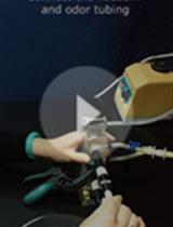
Procedure
文章信息
版权信息
© 2020 The Authors; exclusive licensee Bio-protocol LLC.
如何引用
Readers should cite both the Bio-protocol article and the original research article where this protocol was used:
- Shaw, H. S., Larkin, J. and Rao, Y. (2020). Phototactic T-maze Behavioral Assay for Comparing the Functionality of Color-sensitive Photoreceptor Subtypes in the Drosophila Visual System. Bio-protocol 10(6): e3558. DOI: 10.21769/BioProtoc.3558.
- Shaw, H. S., Cameron, S. A., Chang, W. T. and Rao, Y. (2019). The conserved igsf9 protein borderless regulates axonal transport of presynaptic components and color vision in Drosophila. J Neurosci 39(35): 6817-6828.
分类
神经科学 > 感觉和运动系统 > 视网膜
神经科学 > 基础技术 > 高通量筛选
神经科学 > 行为神经科学 > 实验动物模型
您对这篇实验方法有问题吗?
在此处发布您的问题,我们将邀请本文作者来回答。同时,我们会将您的问题发布到Bio-protocol Exchange,以便寻求社区成员的帮助。