

Study of Microbial Extracellular Vesicles: Separation by Density Gradients, Protection Assays and Labelling for Live Tracking
微生物胞外囊泡的研究:密度梯度分离,保护性分析和活体示踪标记
发布: 2020年01月20日第10卷第2期 DOI: 10.21769/BioProtoc.3502 浏览次数: 5891
评审: Kristin L. ShinglerPooja VermaLiwei Chen
参见作者原研究论文
The authors used this protocol in:
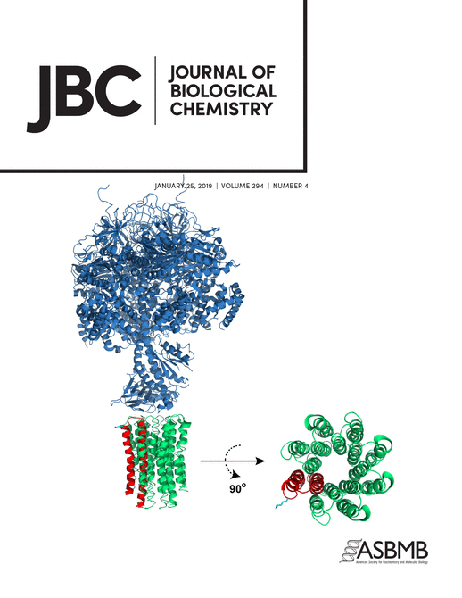
Jan 2019
Advertisement


Abstract
Extracellular vesicles (EVs) are produced by all domains of life including Bacteria, Archaea and Eukarya. EVs are critical for cellular physiology and contain varied cargo: virulence factors, cell wall remodeling enzymes, extracellular matrix components and even nucleic acids and metabolites. While various protocols for isolating EVs have been established for mammalian cells, the field is actively developing tools to study EVs in other organisms. In this protocol we describe our methods to perform density gradient purification of EVs in bacterial cells, allowing for separation of EV subpopulations, followed by protection assays for EV cargo characterization. Furthermore, we devised a protocol which incorporates a fluorescent conjugate of fatty acids into EVs, the first to allow live-cell EV tracking to observe release of EVs, including during infection of mammalian cells by pathogenic bacteria. These protocols are powerful tools for EV researchers as they enable the observation of EV release and the study of the mechanisms of their formation and release.
Keywords: Extracellular Vesicles (胞外囊泡)Background
Microbial extracellular vesicles (EVs) are fundamental for cellular physiology (Deatherage et al., 2012; Brown et al., 2015). For pathogenic microbes, EVs can be harnessed for diagnostics and vaccines. However, the study of EVs has been hampered by limited availability of tools which allow in-depth functional and mechanistic study of these small and extraordinary structures.
The protocol for isolating microbial EVs is well established (Rodrigues et al., 2008) and consists of isolation of culture supernatants followed by several (ultra)-centrifugation cycles to separate EVs from cells and debris. Several other EV isolation techniques have been developed in the last years, but these techniques are usually tested and implemented in the context of mammalian EVs and thus do not readily translate to microbial EVs. Researchers should follow the International Society for Extracellular Vesicles ISEV guidelines for EV isolation (Théry et al., 2018) when implementing such protocols.
In our most recent work (Coelho et al., 2019) we developed a density separation purification, a protection assay and a labelling protocol for the live-cell imaging of EVs from the pathogenic bacteria Listeria monocytogenes. In the protocols described here, growth conditions and media are optimized for L. monocytogenes, but our protocols should be readily adaptable to other microbes by adjusting the media and growth conditions. For example, density gradient purification was first developed for the pathogenic yeast Cryptococcus neoformans (Vij et al., 2018).
In this work we successfully used a multi-omics approach, a single-sample analysis of metabolite, protein, and lipid extraction (MPLEx) (Nakayasu et al., 2016) which provided a very cost effective analysis of EV composition. The MPLEx protocol has been well described elsewhere (Burnum-Johnson et al., 2017; Nicora et al., 2018).
Density separation methods for EVs have been used previously (Horstman and Kuehn, 2000; Elluri et al., 2014) and we adapted it to our specific needs. Density separation methods have two advantages over the use of EV preparations described in Protocol A: i) it allows the study the several subpopulations of EVs and, ii) it increases purity of EV preparations by removing contaminants such as soluble and aggregated proteins. Density separation of EVs extracted from 1 L microbial supernatant from L. monocytogenes (or C. neoformans) generates sufficient EVs for electron microscopy as well as dot blot protocols and we are confident that other analysis, such as proteomics and other -omics, can be performed. We have used variations of this protocol to separate melanin from the pathogenic yeast Cryptococcus neoformans (Camacho et al., 2019). Optiprep was used to prepare a step-density gradient, as we found it more straightforward to remove Optiprep from vesicle samples than other reagents used to prepare density gradients. In our experience, colloidal reagents, such as Percoll, were difficult to remove from preparations and subsequently interfered in downstream analysis of EVs by electron microscopy and western blotting.
Protection Assays in EVs have been performed before by our group (Rodrigues et al., 2008; Brown et al., 2014) as they allow to distinguish EV-internalized cargo from exposed cargo. Protection assays are considered the gold standard to show internalization of cargo versus adsorption or co-purification, and no other method (such as immunostaining and imaging by electron microscopy) is accepted as proof that cargo is internalized. Protection assays consist of subjecting the EV preparation to the action of degradative enzyme and then testing for degradation of EV components. In the specific case of L. monocytogenes, EVs were exposed to a protease (proteinase K or trypsin) that will digest any proteins external to EVs while proteins internalized in EVs are protected from proteases. Since L. monocytogenes secreted listeriolysin O (LLO) in EVs and this virulence factor is very efficient in lysing red blood cells we tested internalization of LLO in EVs by measuring lysis of red blood cells. Variations of these strategies simply use different degradative enzymes (proteases, nucleases, etc.) and test for protection inside EVs by measuring remaining amount of degraded product via immunoblot, activity assays, direct quantification, etc. The key factors for protection assays are gentle handling of the EVs to maintain their integrity, as well as appropriate experimental controls (the EVs from genetic-deleted strains and exogenously added controls such as recombinant proteins).
EV labelling in microbes has proven difficult. Despite wide usage of the lipophilic dyes DiO, DiL and PKH26, in our hands we noticed strong aggregation of EVs after labelling with DiO and DiL dyes (measured via dynamic light scattering). Other authors have reported similar problems (Morales-Kastresana et al., 2017; Dehghani et al., 2019) and we thus strongly discourage the use of DiO, DiL and PKH26 for the study of EVs. Alternatively, authors have used molecular tagging of EV cargo coupled to fluorescent or luminescent proteins. Those approaches are very successful when the protein cargo of EVs has been established, and if the cargo is amenable to molecular tagging. We developed a cargo-independent protocol which allows for live-tracking of bacteria, and EVs released by the bacteria, without altering the size (measured via dynamic light scattering) of EVs (Coelho et al., 2019). In brief, we allowed bacterial membranes to incorporate a conjugate of the fluorescent dye Bodipy558 to a long-chain fatty acid dodecanoic acid (BODIPY® FL C12, Thermo Fisher Scientific) followed by high-resolution live-cell microscopy, allowing us to capture videos of release of EVs (Coelho et al., 2019). The strength of this approach is it can be easily applied to other organisms, from mammalian cells to bacteria and fungi, as other fatty acids-BODIPY dye conjugates exist (hexadecanoic for example). In addition, these conjugates are relatively cheap. For our particular labelling protocol we noted strong staining of bacterial cells as well as EVs. We reason that labelling with other fatty acids conjugates may allow more specific staining of EVs. We would recommend to readers to attempt labelling with other fatty acids- BODIPY conjugates as they optimize for their microbes of choice.
Materials and Reagents
- 96-well Flat Bottom Plate (Millipore Sigma, catalog number: CLS3997)
- 96-well Round Bottom Plate (Millipore Sigma, catalog number: CLS3999)
- 8-well microscopy µ-slides, tissue culture treated (ibidi, catalog number: 80826-90)
- Steritop Vacuum Filter Sterilizer, 0.22 µm, GP Millipore Express Plus Membrane, 150 ml Funnel, 45 mm neck size (Millipore, catalog number: SCGPT01RE)
- Amicon Ultra-4 Centrifugal Filter Unit, 10 kDa (Millipore, catalog number: UFC801096) for buffer exchange
- Pipette tips (USA Scientific TipOne, catalog number: 1111-0006)
- 1.5 ml Eppendorf tube
- 3 ml ultracentrifuge tube
- 100% Sheep Red Blood Cells (Store at 2-8 °C for up to one month, Innovative Research, IC100-0210-26568)
- Human epithelial MCF-7 breast cancer cells (ATCC, catalog number: HTB-22)
- 70% ethanol
- Brain-Heart Infusion (BHI) broth (Gibco, Fisher Scientific, catalog number: 211059) and agar (Fisher Scientific, catalog number: B11065)
- Vegitone Infusion broth (Millipore, Merck, catalog number: 41960-500G-F)
- Optiprep Density Gradient Medium (Sigma, catalog number: D1556-250ML)
- BODIPYTM FL C12 (4,4-Difluoro-5,7-Dimethyl-4-Bora-3a,4a-Diaza-s-Indacene-3-Dodecanoic Acid) (Molecular Probes, Thermo Fisher Scientific, catalog number: D-3822)
- Bovine Serum Albumin (BSA) Fraction V, pH 7.0 (store at 2-8 °C, MP Biomedicals, catalog number: 810034)
- Dithiothreitol (DTT), use stock dilutions kept at -20 °C for no more than a couple of months (store at -20 °C, Invitrogen, catalog number: 15508-013)
- Recombinant LLO (rLLO) (store at -20°C in small aliquots to avoid freez-thaw cycles, Abcam, catalog number: ab83345)
- Trypsin-EDTA (0.05%), Phenol Red (stored -20 °C for up to 2 years, and prior to use thawed and kept at 2-8 °C, Gibco, Thermo Fischer Scientific, catalog number: 25300054)
- Dulbecco's Modified Eagle Medium (DMEM; Corning, catalog number: 10-013-CV), supplemented with 10% FBS (Millipore Sigma, catalog number: TMS-016-B), GlutaMAX (Thermo Fisher Scientific, catalog number: 35050061), non-essential amino acids (Thermo Fisher Scientific, catalog number: 11140076), and Gibco Antibiotic-Antimycotic (Thermo Fisher Scientific, catalog number: 15240096)
- DMEM without phenol red (Thermo Fisher Scientific, catalog number: 11054020) with 10% FBS (Millipore Sigma, catalog number: TMS-016-B), GlutaMAX (Thermo Fisher Scientific, catalog number: 35050061), and non-essential amino acids (Thermo Fisher Scientific, catalog number: 11140076)
- Hoechst 33342 (1 µg/ml; Thermo Fisher Scientific, catalog number: H3570)
- TrypLE (Thermo Fisher Scientific, catalog number: 12604013)
- Phosphate buffered saline (PBS), pH 7.4, cell culture grade (Corning, catalog number: 21-031-CV)
- 4% paraformaldehyde (PFA; Electron Microscopy Sciences, catalog number: 15710)
- Gentamicin (Thermo Fisher Scientific, catalog number: 15710064)
- 0.02% sodium azide (Sigma, catalog number: S2002-5G)
- HCl (Fisher Scientific, catalog number: A144-212)
- HEPES (Sigma, catalog number: H3375)
- NaCl (Fisher Scientific, catalog number: S271)
- DMSO (Sigma, catalog number: D2650)
- RBC Lysis buffer (see Recipes)
Equipment
- Ultracentrifuge (Beckman, model: Optima-XL) with SW Ti 55 rotor
- Sorvall Centrifuge (Sorvall RC 5C Plus)
- Sorvall GSA fixed-angle rotor
- Polycarbonate Ultracentrifuge Tubes, 3 ml (Beckman Coulter, catalog number: 355635)
- Ultra-Clear Tube, 25 x 89mm, 38.5 ml (Beckman Coulter, catalog number: 344058)
- Multi-Angle Particle Sizing analyzer (Brookhaven Instruments Corp, 90Plus/ BI-MAS)
- Microbiology shaking incubator, heated water bath
- For EV isolation of large volumes cultures, ultrafiltration or centrifugation devices [Amicon Stirred Cell Model 8200, 200 ml (Millipore, catalog number: WW-29968-16) with Ultracel 100 kDa Ultrafiltration Disks (Millipore, catalog number: PLHK07610) with a N2 source]
- Nalgene 250 ml Centrifuge bottles (Thermo Fischer Scientific, catalog number: 3120-0250)
- Multichannel pipettes (BrandTech Scientific Inc., Transferpette S-12 Multichannel Pipettes, catalog number: 703730)
- Dot blot apparatus (such as Bio-Dot® Microfiltration System (Bio-Rad, catalog number: 1703938) or apply samples to membrane manually)
- DeltaVision Elite High-Resolution Deconvolution Microscope (GE Healthcare) equipped with:
- A Scientific CMOS camera (Chip size: 2560 x 2160 pixels)
- An UltraFast solid-state illumination
- An environmental chamber for live cell imaging
- A 60x (N.A. 1.42) oil immersion objective
- UltimateFocus module
Software
- SoftWoRx (Applied Precision, www.gelifesciences.com)
- ImageJ/FIJI (http://fiji.sc/)
Procedure
文章信息
版权信息
© 2020 The Authors; exclusive licensee Bio-protocol LLC.
如何引用
Readers should cite both the Bio-protocol article and the original research article where this protocol was used:
- Coelho, C., Vij, R., Smith, D. Q., Brady, N. R., Hamacher-Brady, A. and Casadevall, A. (2020). Study of Microbial Extracellular Vesicles: Separation by Density Gradients, Protection Assays and Labelling for Live Tracking. Bio-protocol 10(2): e3502. DOI: 10.21769/BioProtoc.3502.
- Coelho, C., Brown, L., Maryam, M., Vij, R., Smith, D. F. Q., Burnet, M. C., Kyle, J. E., Heyman, H. M., Ramirez, J., Prados-Rosales, R., Lauvau, G., Nakayasu, E. S., Brady, N. R., Hamacher-Brady, A., Coppens, I. and Casadevall, A. (2019). Listeria monocytogenes virulence factors, including listeriolysin O, are secreted in biologically active extracellular vesicles. J Biol Chem 294(4): 1202-1217.
分类
微生物学 > 微生物-宿主相互作用 > 细菌
微生物学 > 微生物细胞生物学 > 细胞成像
细胞生物学 > 细胞器分离 > 胞外囊泡
您对这篇实验方法有问题吗?
在此处发布您的问题,我们将邀请本文作者来回答。同时,我们会将您的问题发布到Bio-protocol Exchange,以便寻求社区成员的帮助。