

A Simple and Efficient Method for Concomitant Isolation and Culture of Enriched Astroglial and Microglial Cells from the Rat Spinal Cord
一种简单有效的大鼠脊髓星形胶质细胞和小胶质细胞联合分离培养方法
发布: 2020年01月20日第10卷第2期 DOI: 10.21769/BioProtoc.3501 浏览次数: 5160
评审: LAXMI NARAYAN MISHRAFanny EhretAlexandros C Kokotos
参见作者原研究论文
The authors used this protocol in:

Dec 2017
Advertisement


Abstract
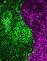
Investigations into glial biology have contributed substantially in understanding the physiology and pathology of the nervous system. However, intricacies of the neuron-glial and glial-glial interactions in vivo present significant challenges while delineating the individual cell-type contributions, thus making the in vitro techniques exceedingly relevant to study glial biology. However, obtaining optimal yield along with high purity has been challenging for microglial cultures. Here we present a simple protocol to establish enriched astroglial as well as microglial cultures from the neonatal rat spinal cord. This method results in highly enriched astroglial and microglial cultures with maximal yield.
Keywords: Primary cultures (原代培养)Background
Astrocytes and microglial cells play a crucial role during the development, as well as in the normal physiology and pathology of the nervous system. These include structural, vascular, metabolic and neuro-regulatory functions like modulation of transmission by forming ‘quad-partite synapses’, release of gliotransmitters and trophic factors, and integration of neuronal-glial networks (Parpura et al., 1994; Araque et al., 1999; Schafer et al., 2013; Michell-Robinson et al., 2015; Rossi, 2015). Microglia, in addition to the canonical immune functions, also plays an important role in adult neurogenesis, differentiation, maturation and integration within the neuronal circuitry (Rezaie and Male, 2002; Polazzi and Monti, 2010; Michell-Robinson et al., 2015; Ransohoff and El Khoury, 2015). In response to a pathological insult, the glial cells react by undergoing morphological and physiological changes and rescue the vulnerable neurons from the insult (Ferraiuolo et al., 2011; Pekny et al., 2014; Mishra et al., 2016; Mishra et al., 2017). At the tissue level, astrocytes undergo morphological transformation from protoplasmic to fibrillary phenotype and move to the site of injury to create a physical barrier between damaged and healthy cells, or by forming glial scars in a process called reactive gliosis. In a similar manner, microglia undergo morphological transformation from the resting/ramified phenotype to the activated/amoeboid phenotype, via intermediate activation stages, (Rezaie and Male, 2002; Michell-Robinson et al., 2015; Ransohoff and El Khoury, 2015). Both the glial cell types thus promote neuronal repair through acute inflammation, which modulates the microenvironment by regulating production and release of trophic factors, gliotransmitters, cytokines and chemokines (Raivich et al., 1999; Parpura et al., 2012; Verkhratsky and Butt, 2013; Pekny et al., 2014). Reactive gliosis is reversible and beneficial during the acute insult. However, in response to prolonged insult or genetic dysregulation, the balance is disrupted and may lead to irreversible and erroneous activation resulting in chronic inflammation and neurodegeneration (Lobsiger and Cleveland, 2007). Therefore, delineating the precise glial response as well as their role in modulating neuronal physiology becomes relevant. However, an in vivo approach may present with a disadvantage owing to the complex neuron-glial and glial-glial interactions, which dynamically modulate these changes at the tissue level. Primary glial cultures help us to overcome this issue, by enabling observations in an isolated cell-type model system. In vitro experimentation also widens the scope for further comparing the effect of neuron-glial/glial-glial interactions through a co-culture model system.
Various approaches have been adopted to culture astrocytes and microglia from animal brains and spinal cords. These methods vary considerably not only in terms of purity and yield, but also with respect to the cell population targeted for isolation (Giulian and Baker, 1986; Saura et al., 2003; Floden and Combs, 2007; Scorisa et al., 2010; Kerstetter and Miller, 2012). For instance, the conventional repeated shaking methods for culturing microglia exploit the microglial layer growing atop astrocytes in the mixed glial cultures (Giulian and Baker, 1986; Scorisa et al., 2010), while the mild trypsinization method targets the ones growing beneath (Saura et al., 2003). We compared all the methods and standardized a procedure to establish exceedingly enriched astroglial and microglial cultures, while maximizing the yield. The current protocol generates enough microglial and astroglial cells per spinal cord to execute parallel experiments in both the cell types, thus minimizing the inter-population variability while comparing cell-specific responses.
Materials and Reagents
- Animals
Wistar strain rat pups with post-natal Day 0-2 (P0-P2), housed and maintained in accordance with the institutional ethics guidelines - Culture products
- 0.22 µm membrane filter (Millipore, catalog number: GSWP04700)
- T-25 ml tissue culture flask (Corning, Sigma, catalog number: CLS430639)
- 24-well plate (Thermo Fisher Scientific, catalog number: 142475)
- Coverslips, 13 mm, circular (Thermo Fisher Scientific, catalog number: 12-519-21G)
- DNase-1 (Sigma-Aldrich, catalog number: 11 284 932 001)
- Hanks’ balanced salt solution (HCMF, Thermo Fisher Scientific, catalog number: 14170112)
- Poly-L-lysine hydrobromide (PLL, Sigma-Aldrich, catalog number: P5899-5G)
- Dulbecco’s modified Eagle’s medium/F-12 (DMEM/F-12) (GIBCO, Invitrogen, USA, catalog number: 12500062)
- L-Glutamine, 200 mM solution (Thermo Fisher Scientific, catalog number: 25030081)
- Fetal bovine serum-Heat inactivated (FBS-HI) (GIBCO, Invitrogen, USA, catalog number: 10082147)
- 0.25% trypsin-EDTA (GIBCO, Invitrogen, USA, catalog number: 25200)
- Penicillin-streptomycin (Pen/Strep; 100x solution; 10,000 units/ml each) (Thermo Fisher Scientific, catalog number: 15140122)
- HEPES (1 M) (Thermo Fisher Scientific, catalog number: 15630080)
- MilliQ water
- HCl (Sigma-Aldrich, catalog number: 7647-01-0)
- Ethanol (Sigma-Aldrich, catalog number: 64-17-5)
- HBSS
- Glucose powder
- NaCl
- KCl
- Na2HPO4
- KH2PO4
- PFA
- Growth medium (500 ml) (see Recipes)
- 50 ml FBS-HI (10% v/v) (see Recipes)
- Dissection medium (500 ml) (see Recipes)
- Dissociation medium (10 ml) (see Recipes)
- DMEM-trypsin-EDTA (1:4) (10 ml) (see Recipes)
- Immunostaining
- 0.1 M PBS, freshly prepared
- Bovine serum albumin (BSA) (Sigma-Aldrich, catalog number: A2153)
- Anti-ChAT (rabbit polyclonal Abcam, catalog number: ab18736; 1:500)
- Anti-GFAP (mouse monoclonal, Abcam, catalog number: ab24345; 1:500)
- Anti-IBA1 (Rabbit polyclonal Abcam, catalog number: ab153696; 1:500)
- Anti-mouse IgG (Cy3-conjugated; 1:200, Sigma-Aldrich)
- Anti-rabbit IgG (FITC-conjugated; 1:200, Chemicon)
- PVA-DABCO anti-fade mounting medium (Sigma-Aldrich, catalog number: 10981)
- PBS 1x (pH-7.4) (see Recipes)
- PFA, 4% (see Recipes)
Equipment
- Fine forceps, Dumont #5 (2), curved (1) (Fine Science Tools)
- Scissors (1), 51/2 in., straight, operating (Fine Science Tools)
- Scissors (2), 4 in., straight, micro dissecting (Fine Science Tools)
- Dissecting microscope (Leica Microsystems)
- Phase contrast microscope (Leica Microsystems)
- Confocal laser-scanning microscope (Leica Microsystems)
- CO2 incubator (Eppendorf)
- Orbital Shaker (New Brunswick, Eppendorf)
- Centrifuge (Thermo Scientific)
- Fire polished glass pipette (Fisherbrand)
- Light source
- Laminar flow hood
- 4 °C refrigerator
- -20 °C freezer
- -80 °C freezer
- Hemocytometer
Software
- Photoshop CS2 (Adobe)
- Microsoft Office
Procedure
文章信息
版权信息
© 2020 The Authors; exclusive licensee Bio-protocol LLC.
如何引用
Mishra, P. S. and Raju, T. R. (2020). A Simple and Efficient Method for Concomitant Isolation and Culture of Enriched Astroglial and Microglial Cells from the Rat Spinal Cord. Bio-protocol 10(2): e3501. DOI: 10.21769/BioProtoc.3501.
分类
神经科学 > 细胞机理 > 细胞分离和培养
细胞生物学 > 细胞分离和培养 > 单层培养
您对这篇实验方法有问题吗?
在此处发布您的问题,我们将邀请本文作者来回答。同时,我们会将您的问题发布到Bio-protocol Exchange,以便寻求社区成员的帮助。