- EN - English
- CN - 中文
In vitro Differentiation of Human iPSC-derived Retinal Pigment Epithelium Cells (iPSC-RPE)
人诱导多功能干细胞来源的视网膜色素上皮细胞的体外分化
发布: 2019年12月20日第9卷第24期 DOI: 10.21769/BioProtoc.3469 浏览次数: 7910
评审: Giusy TornilloTomohiro MizutaniAgnieszka Maria Pastula

相关实验方案

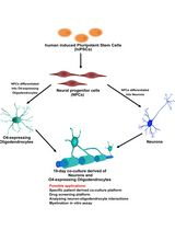
人 iPSC 衍生神经元与少突胶质细胞共培养用于髓鞘形成的小分子筛选分析
Stefanie Elke Chie [...] Maria Consolata Miletta
2025年05月05日 3259 阅读
Abstract
Induced Pluripotent Stem Cells (iPSCs) serve as an excellent model system for studying the molecular underpinnings of tissue development. Human iPSC-derived retinal pigment epithelium (iPSC-RPE) cells have fetal-like molecular profiles. Hence, biobanks like iPSCORE, which contain iPSCs generated from hundreds of individuals, are an invaluable resource for examining how common genetic variants exert their effects during RPE development resulting in individuals having different propensities to develop Age-related Macular Degeneration (AMD) as adults. Here, we present an optimized, cost-effective and highly reproducible protocol for derivation of human iPSC-RPE cells using small molecules under serum-free condition and for their quality control using flow cytometry and immunofluorescence. While most previous protocols have required laborious manual selection to enrich for iPSC-RPE cells, our protocol uses whole culture passaging and yields a large number of iPSC-RPE cells with high purity (88-98.1% ZO-1 and MiTF double positive cells). The simplicity and robustness of this protocol would enable its adaption for high-throughput applications involving the generation of iPSC-RPE samples from hundreds of individuals.
Keywords: Human induced pluripotent stem (hiPSC) (人诱导多功能干细胞)Background
Age-related macular degeneration (AMD) is a leading cause of vision loss in developed countries affecting 11 million individuals in the United States and about 170 million worldwide (Pennington and DeAngelis, 2016). Moreover, considering age as a main factor, in our current aging society, the incidence of AMD is estimated to increase to 198 million in 2020 and 288 million by 2040 and to 22 million in the United States alone by 2050 (Wong et al., 2014, Pennington and DeAngelis, 2016). Current therapeutic strategies, although effective, are expensive and limited to delaying the speed of disease progression, and AMD still eventually leads to a complete loss of vision (Al-Zamil and Yassin, 2017; Mitchell et al., 2018). At the time of preparation of this article, there are seven active clinical trials aimed to evaluate the effectiveness and optimize the conditions of the transplantation of the human iPSC-RPEs or human embryonic stem cell-derived retinal pigment epithelium (ESC-RPE) cells (NIH-ClinicalTrials.gov). Thus, the development of a robust and cost-effective method for generating large amounts of high quality iPSC-RPEs is imperative for the advancement of future therapeutic treatments of AMD and potentially other eye diseases in humans as well as domestic animals (Sparrow et al., 2010).
We have previously demonstrated the utility of employing iPSC-RPE cells to identify and study genetic variants playing a role in the development of AMD (Smith et al., 2019). In this study, we derived iPSC-RPE cells from six individuals (3 European Americans, 2 East Asian Americans, and 1 African American) and showed that they have morphological and molecular characteristics similar to those of naïve RPE cells. We showed that iPSC-RPE gene expression profiles are highly similar to that of human fetal RPE, and that their ATAC-seq (Assay for Transposase-Accessible Chromatin using sequencing) peaks are enriched for relevant transcription factor motifs. We performed fine mapping of AMD risk loci integrating the molecular data from iPSC-RPE cells that resulted in the prioritization of four variants, including a potential regulatory SNP (rs943080) near VEGFA and a coding SNP (rs34882957, pP167S) in the C9 gene (Smith et al., 2019). Our findings illustrate that iPSC-RPE cells are an excellent model system to study the molecular functions of genetic variation associated with AMD.
The initial protocols for deriving iPSC-RPE cells developed over a decade ago involved spontaneous differentiation and were highly inefficient requiring the manual separation of pigmented iPSC-RPE patches from non-differentiated cells (Klimanskaya et al., 2004; Vugle et al., 2008). Subsequently, induced directed differentiation protocols resulted in higher yielded and quality of iPSC-RPE cells but required numerous animal-derived components and thus had relatively low reproducibility or were very time consuming (Aoki et al., 2006; Osakada et al., 2008; Buchholz et al., 2009; Idelson et al., 2009; Reichman et al., 2014; Hazim et al., 2017). The introduction of small molecules into iPSC-RPE differentiation protocols greatly simplified the procedure and resulted in higher reproducibility (Osakada et al., 2009; Maruotti et al., 2013; Maruotti et al., 2015). Previous studies have optimized differentiation protocols to derive iPSC-RPEs from a limited number of iPSC or ESC lines and in most cases utilized small format culture vessels. Here, we present an optimized protocol for deriving iPSC-RPEs from multiple human iPSC lines in large sized culture flasks. It is similar to the protocol of Maruotti et al. (2015), but initiates differentiation when iPSCs reach 80% confluency, which we found to be optimal for all tested iPSCORE lines (Panopoulos et al., 2017). Additionally, we modified the length of time of exposure to small molecules at two steps, which resulted in further increase of yield and purity of iPSC-RPEs (88-98.1% ZO-1 and MiTF double positive cells). Our optimized protocol allowed us to derive iPSC-RPEs from six iPSC lines generated from ethnically diverse individuals under identical culturing conditions without the requirement of any individualized optimization steps.
Materials and Reagents
- iPSC Cell Culture
- 6-well plates (Corning, catalog number: 3506)
- Syringe filter 0.2 μm (VWR, catalog number: 28145-501)
- Soft-Ject® 3-Part Disposable Syringe, Air-Tite-3 ml (VWR, catalog number: 89215-234)
- 5 ml Borosilicate serological pipettes (Fisher Scientific, catalog number: 1367827E)
- 5 ml Serological pipettes (Bio Pioneer, catalog number: GEX0050-S01)
- 10 ml Serological pipettes (Bio Pioneer, catalog number: GEX0100-S01)
- P20 pipette tips sterile with filter
- P1000 pipette tips sterile with filter
- 15 ml conical tubes (Bio Pioneer, catalog number: CNT-15R)
- iPSC cells
- 70% ethanol
- UltraPureTM DNase/RNase-Free Distilled Water (Thermo Fisher Scientific, catalog number: 10977023)
- Corning® Matrigel® Growth Factor Reduced (GFR) Basement Membrane Matrix (Corning, catalog number: 354230)
- mTeSRTM1 (Stem Cell Technologies, catalog number: 85850)
- DMEM/F-12 medium (Thermo Fisher Scientific, catalog number: 11330057)
- Dispase II (Thermo Fisher Scientific, catalog number: 17105041)
- Matrigel solution (Matrigel) (see Recipes: Table 1)
- 10 mM ROCK inhibitor, Y-27632 dihydrochloride solution (ROCK Inhibitor) (see Recipes: Table 2)
- 10x Dispase (see Recipes: Table 3)
- mTeSRTM1 complete medium (mTeSR) (see Recipes: Table 4)
- Monolayer plating
- 100 mm tissue culture dishes (Corning, catalog number: 430167)
- Automated cell counter slides (Bio-Rad Laboratories, catalog number: 1450019) or a hemocytometer (Hausser Scientific, catalog number: 1483) or equivalent
- 5 ml Serological pipettes (Bio Pioneer, catalog number: GEX0050-S01)
- 10 ml Serological pipettes (Bio Pioneer, catalog number: GEX0100-S01
- P20 pipette tips sterile with filter
- P200 pipette tips sterile with filter
- P1000 pipette tips sterile with filter
- 15 ml conical tubes (Bio Pioneer, catalog number: CNT-15R)
- 50 ml conical tubes (Bio Pioneer catalog number: CNT-50R)
- 70% ethanol
- Corning® Matrigel® Growth Factor Reduced (GFR) Basement Membrane Matrix (Matrigel) (Corning, catalog number: 354230)
- mTeSRTM1 (Stem Cell Technologies, catalog number: 85850)
- DMEM/F-12 medium (Thermo Fisher Scientific, catalog number: 11330-057)
- Accutase (Innovative Cell Technologies, Inc., catalog number: AT 104)
- Trypan Blue Solution, 0.4% (Thermo Fisher Scientific, catalog number: 15250061)
- ROCK inhibitor, Y-27632 dihydrochloride (Selleck hem, catalog number: S1049)
- iPSC cell culture
- Matrigel solution (see Recipes: Table 1)
- 10 mM ROCK inhibitor, Y-27632 dihydrochloride solution (see Recipes: Table 2)
- mTeSRTM1 complete medium (see Recipes: Table 4)
- iPSC-RPE differentiation
- 100 mm tissue culture dishes (Corning, catalog number: 430167)
- (Optional) T150 tissue culture flasks, vented (Sigma, catalog number: Z707929)
Note: At the time of preparation of this manuscript Z707929 was no longer available. The same flasks are available under the catalog number Z707511-36EA (Sigma, catalog number Z707511-36EA). - 70 μm strainers (Fisher Scientific, catalog number: 431751)
- Automated cell counter slides (Bio-Rad Laboratories, catalog number: 1450019) or a hemocytometer (Hausser Scientific, catalog number: 1483) or equivalent
- 10 ml Serological pipettes (Bio Pioneer, catalog number: GEX0100-S01)
- 25 ml Serological pipettes (Bio Pioneer catalog number: GEX250-S01)
- 50 ml Serological pipettes (Bio Pioneer, catalog number: GEX500-S01)
- P20 pipette tips sterile with filter
- P200 pipette tips sterile with filter
- P1000 pipette tips sterile with filter
- Cell scraper (VWR International, catalog number: 179707)
- 15 ml conical tubes (Bio Pioneer, catalog number: CNT-15R)
- 50 ml conical tubes (Bio Pioneer, catalog number: CNT-50R)
- Nalgene Cryogenic vials (Thermo Fisher Scientific, catalog number: 5000-1020)
- iPSCs monolayer
- 70% ethanol
- Corning® Matrigel® Growth Factor Reduced (GFR) Basement Membrane Matrix (Matrigel) (Corning, catalog number: 354230)
- DMEM/F-12 medium (Thermo Fisher Scientific, catalog number: 11330057)
- DMEM medium (Thermo Fisher Scientific, catalog number: 11965092)
- Ham’s F12 Nutrient Mix (Thermo Fisher Scientific, catalog number: 11765054)
- 1x Dulbecco’s phosphate buffered saline (DPBS) without calcium and magnesium (Thermo Fisher Scientific, catalog number: 14190250)
- B27 Supplement (50x), serum free (Thermo Fisher Scientific, catalog number: 17504044)
- KnockOutTM Serum Replacement (KOSR) (Thermo Fisher Scientific, catalog number: 10828028)
- L-Glutamine 200 mM (Thermo Fisher Scientific, catalog number: 25030081)
- MEM Non-Essential Amino Acids Solution 100x (Thermo Fisher Scientific, catalog number: 11140050)
- Penicillin-Streptomycin (10,000 U/ml) (Thermo Fisher Scientific, catalog number: 15140122)
- β-Mercaptoethanol (Thermo Fisher Scientific, catalog number: 21985023)
- Accutase (Innovative Cell Technologies, Inc., catalog number: AT 104)
- Nicotinamide (Sigma, catalog number: N3376)
- Chetomin (Sigma, catalog number: C9623)
- Trypan Blue Solution, 0.4% (Thermo Fisher Scientific, catalog number: 15250061)
- Dimethyl Sulfoxide (DMSO) (Sigma, catalog number: D2650-100ML)
- Liquid nitrogen
- RPE DM medium (see Recipes: Table 5)
- RPE medium (see Recipes: Table 6)
- 2x iPSC-RPE freezing medium (see Recipes: Table 7)
- 1 mM Chetomin solution (see Recipes: Table 8)
- 1 M Nicotinamide (100x) solution (see Recipes: Table 9)
- Flow cytometry
- 96-well round bottom assay plates (Genesee Scientific, catalog number: 25-224)
- CorningTM FalconTM Test Tube with Cell Strainer Snap Cap (Fisher Scientific, catalog number: 352235)
- CorningTM CostarTM Sterile Disposable Reagent Reservoirs (Fisher Scientific, catalog number: 4870)
- 5 ml Serological pipettes (Bio Pioneer, catalog number: GEX0050-S01)
- 10 ml Serological pipettes (Bio Pioneer, catalog number: GEX0100-S01)
- P20 pipette tips sterile with filter
- P200 pipette tips sterile with filter
- P1000 pipette tips sterile with filter
- P20 pipette tips without filter
- (Optional) P200 pipette tips without filter
- Fixation/Permeabilization Solution Kit with BD GolgiStopTM (BD Biosciences, catalog number: 554715)
- 1x Dulbecco’s phosphate buffered saline (DPBS) without calcium and magnesium (Thermo Fisher Scientific, catalog number: 14190250)
- Bovine Serum Albumin (BSA) (Sigma, catalog number: A2153-100G)
- (Optional) NaN3 (Sigma, catalog number: S2002-5G)
- 37% Formaldehyde (Sigma, catalog number: F-1635-500ML)
- Rabbit polyclonal anti-ZO-1 antibody (abcam, catalog number: ab59720)
Note: During preparation of this manuscript the antibody ab59720 was no longer available. A potential replacement: abcam, catalog number: ab221547. - Mouse monoclonal anti-MiTF antibody (abcam, catalog number: ab12039)
- Recombinant Rabbit IgG, monoclonal [EPR25A]–Isotype Control (abcam, catalog number: ab172730)
- Mouse IgG1, kappa monoclonal [15-6E10A7]–Isotype Control (abcam, catalog number: ab170190)
- Donkey-anti-Rabbit Alexa FluorTM 647 conjugated antibody (abcam, catalog number: ab150075)
- Goat-anti-Mouse Alexa FluorTM 488 conjugated antibody (Thermo Scientific, catalog number: A-11001)
- FACS Buffer (see Recipes: Table 10)
- FACS-FIX Buffer (see Recipes: Table 11)
- Immunofluorescence
- Millicell EZ SLIDE 8-well glass slides (Millipore, catalog number: PEZGS0816)
- Cover glass slides (Fisherbrand, catalog number: 12-545-F)
- 5 ml Serological pipettes (Bio Pioneer, catalog number: GEX0050-S01)
- 10 ml Serological pipettes (Bio Pioneer, catalog number: GEX0100-S01)
- P20 pipette tips sterile with filter
- P200 pipette tips sterile with filter
- 1x Dulbecco’s phosphate buffered saline (DPBS) without calcium and magnesium (Thermo Fisher Scientific, catalog number: 14190250)
- Bovine Serum Albumin (BSA) (Sigma, catalog number: A2153-100G)
- Paraformaldehyde (PFA)
- Tween® 20 (Sigma, catalog number: P9416-100ML)
- Triton X-100 (Manufacturer, catalog number: X-100-500ML)
- Corning® Matrigel® Growth Factor Reduced (GFR) Basement Membrane Matrix (Matrigel) (Corning, catalog number: 354230)
- Rabbit polyclonal anti-ZO-1 antibody (abcam, catalog number: ab59720)
- Mouse monoclonal anti-MiTF antibody (abcam, catalog number: ab12039)
- Mouse monoclonal anti-Bestrophin 1 antibody (Novus Biologicals, catalog number: NB300-164SS)
- Recombinant Rabbit IgG, monoclonal [EPR25A]–Isotype Control (abcam, catalog number: ab172730)
- Mouse IgG1, kappa monoclonal [15-6E10A7]–Isotype Control (abcam, catalog number: ab170190)
- Donkey-anti-Rabbit Alexa FluorTM 647 conjugated antibody (abcam, catalog number: ab150075)
- Goat-anti-Mouse Alexa FluorTM 488 conjugated antibody (Thermo Scientific, catalog number: A-11001)
- ProLong Gold Antifade Reagent with DAPI (Cell Signaling Technologies, catalog number: 8961)
- IF Wash Buffer (see Recipes: Table 12)
- IF Perm Buffer (see Recipes: Table 13)
- IF Staining Buffer (see Recipes: Table 14)
Equipment
- iPSC Cell Culture
- Biosafety cabinet (Labconco, model: Logic+)
- Incubator with humidity and gas control set to maintain 37 °C and 95% humidity in an atmosphere of 5% CO2 in air (Panasonic, model: MCO-170AICUVH-PA)
- Water bath (Thermo Scientific, model: Precision)
- Tissue culture centrifuge with rotors for 15 ml conical tubes and 50 ml conical tubes (Thermo Scientific, model: Legend RT+)
- Phase contrast inverted microscope (objectives: x4, x10, x20) (Olympus, model: CKX41SF)
- (Optional) Phase contrast inverted microscope with camera (objectives: x4, x10, x20) (Thermo Scientific, model: EVOS XL Core)
- Microscope Object marker (Nikon, model MBW10020)
- Pipette aid
- P20 Micropipette
- Freezer -20 °C
- Refrigerator 2-8 °C
- Monolayer plating
- Biosafety cabinet (Labconco, model: Logic+)
- Incubator with humidity and gas control set to maintain 37 °C and 95% humidity in an atmosphere of 5% CO2 in air (Panasonic, model: MCO-170AICUVH-PA)
- Tissue culture centrifuge with rotors for 15 ml conical tubes and 50 ml conical tubes (Thermo Scientific, model: Legend RT+)
- Phase contrast inverted microscope (objectives: x4, x10, x20) (Olympus, model: CKX41SF)
- Phase contrast inverted microscope with camera (objectives: x4, x10, x20) (Thermo Scientific, model: EVOS XL Core)–Optional
- Pipette aid
- P20 Micropipette
- P200 Micropipette
- P1000 Micropipette
- Automated cell counter (Bio-Rad, model: TC20) or a hemocytometer (Hausser Scientific, catalog number: 1483) or equivalent
- Freezer -20 °C
- Refrigerator 2-8 °C
- iPSC-RPE differentiation and cryopreservation
- Biosafety cabinet (Labconco, model: Logic+)
- Incubator with humidity and gas control set to maintain 37 °C and 95% humidity in an atmosphere of 5% CO2 in air (Panasonic, model: MCO-170AICUVH-PA)
- Tissue culture centrifuge with rotors for 15 ml conical tubes and 50 ml conical tubes (Thermo Scientific, model: Legend RT+)
- Phase contrast inverted microscope (objectives: x4, x10, x20) (Olympus, model: CKX41SF)
- (Optional) Phase contrast inverted microscope with camera (objectives: x4, x10, x20) (Thermo Scientific, model: EVOS XL Core)
- Pipette aid
- P20 Micropipette
- P200 Micropipette
- P1000 Micropipette
- Automated cell counter (Bio-Rad, model: TC20) or a hemocytometer (Hausser Scientific, catalog number: 1483) or equivalent.
- Mr. Frosty freezing container (Corning, model: CoolCell® FTS30)
- Refrigerator 2-8 °C
- Freezer -20 °C
- Freezer -80 °C
- Liquid nitrogen vapor tank
- Flow cytometry
- Pipette aid
- P20 Micropipette
- P200 Micropipette
- P1000 Micropipette
- P200 Multichannel micropipette
- Refrigerator 2-8 °C
- Freezer -20 °C
- Flow cytometer (BD Biosciences, model: FACSCanto II) or equivalent
- Immunofluorescence
- Pipette aid
- P20 Micropipette
- P200 Micropipette
- P1000 Micropipette
- Refrigerator 2-8 °C
- Freezer -20 °C
- Confocal laser scanning fluorescence microscope (Olympus, FluoView1000)
Software
- FlowJo (Version 10) (FlowJo, LLC, https://www.flowjo.com/)
- FlowView ASW V03.01.03.03 or V4.2a (Olympus Life Science, https://www.olympus-lifescience.com/en/support/downloads/)
Procedure
文章信息
版权信息
© 2019 The Authors; exclusive licensee Bio-protocol LLC.
如何引用
D’Antonio-Chronowska, A., D’Antonio, M. and Frazer, K. A. (2019). In vitro Differentiation of Human iPSC-derived Retinal Pigment Epithelium Cells (iPSC-RPE). Bio-protocol 9(24): e3469. DOI: 10.21769/BioProtoc.3469.
分类
干细胞 > 多能干细胞 > 细胞分化
细胞生物学 > 细胞分离和培养 > 细胞分化
发育生物学 > 细胞生长和命运决定 > 分化
您对这篇实验方法有问题吗?
在此处发布您的问题,我们将邀请本文作者来回答。同时,我们会将您的问题发布到Bio-protocol Exchange,以便寻求社区成员的帮助。
Share
Bluesky
X
Copy link


