

Time-lapse Imaging of Alveologenesis in Mouse Precision-cut Lung Slices
小鼠离体肺组织切片中肺泡生成的延时成像
发布: 2019年10月20日第9卷第20期 DOI: 10.21769/BioProtoc.3403 浏览次数: 7998
评审: Zinan ZhouThirupugal GovindarajanKarthik KrishnamurthyAnita Umesh
参见作者原研究论文
The authors used this protocol in:

Mar 2019
Advertisement


相关实验方案
Abstract
Alveoli are the gas-exchange units of lung. The process of alveolar development, alveologenesis, is regulated by a complex network of signaling pathways that act on various cell types including alveolar type I and II epithelial cells, fibroblasts and the vascular endothelium. Dysregulated alveologenesis results in bronchopulmonary dysplasia in neonates and in adults, disrupted alveolar regeneration is associated with chronic lung diseases including COPD and pulmonary fibrosis. Therefore, visualizing alveologenesis is critical to understand lung homeostasis and for the development of effective therapies for incurable lung diseases. We have developed a technique to visualize alveologenesis in real-time using a combination of widefield microscopy and image deconvolution of precision-cut lung slices. Here, we describe this live imaging technique in step-by-step detail. This time-lapse imaging technique can be used to capture the dynamics of individual cells within tissue slices over a long time period (up to 16 h), with minimal loss of fluorescence or cell toxicity.
Keywords: Imaging alveologenesis (肺泡生成成像)Background
Prenatal and postnatal lung development is classified into several distinct stages beginning with budding from the foregut endoderm followed by branching morphogenesis, sacculation and alveologenesis within surrounding lung mesenchyme. This developmental process is tightly regulated by a well-orchestrated signaling programme and cellular components (Kotton and Morrisey, 2014; Akram et al., 2016). The major function of the lungs is gas exchange, which occurs via diffusion (Herriges and Morrisey, 2014). This diffusion takes place between the thin cellular layers of alveolar epithelium and capillary endothelium (Roth-Kleiner and Post, 2005). Dysregulated alveologenesis is linked with a number of neonatal and infant diseases, including bronchopulmonary dysplasia (BPD) and pulmonary hypoplasia (Kreiger et al., 2006; Hilgendorff et al., 2014). In adults, alveolar damage is a component of several chronic lung diseases such as chronic obstructive pulmonary disease (COPD) and idiopathic pulmonary fibrosis (IPF). Currently, there are no curative treatments for these diseases other than lung transplantation (Warburton et al., 2006; Madurga et al., 2013; McGowan, 2014) and there is an unmet need to understand the mechanisms of alveologenesis in order to develop effective treatments.
In mice, sacculation begins around embryonic day 17.5 and is followed by alveologenesis, which begins within the first few days of postnatal life and is mostly completed in first month of life (Herriges and Morrisey, 2014). However, the most active phase of alveologenesis occurs in first two weeks of postnatal life with the majority of alveoli formed by postnatal day (P) 21 (Hind et al., 2002; Snoeck, 2015).
Current understanding, based on static imaging experiments, is that alveologenesis occurs through repeated septation events that sub-divide primary air sacs to increase the number and surface area of alveoli (Amy et al., 1977; Mund et al., 2008). Real-time visualization of alveologenesis is challenging due to their location deep inside the body and the relatively slow duration of this process. A recent study used both ex vivo and in vivo live-imaging to study the sacculation stage of mouse lung development, immediately prior to alveologenesis, but these techniques are not suitable for imaging postnatal lungs (Poobalasingam et al., 2017; Li et al., 2018).
Precision cut lung slices (PCLS) contain intact alveoli and are increasingly used to study lung biology and disease pathogenesis (Meng et al., 2008; Sanderson, 2011; Thornton et al., 2012). Time-lapse imaging of PCLS has been used to show dynamic interactions of mesenchymal cells and macrophages with the extracellular matrix in adult normal and fibrotic mouse lungs, as well as in PCLS of human lungs (Burgstaller et al., 2015). In addition, quantifiable ex vivo alveologenesis has been demonstrated in early postnatal mouse PCLS culture (Pieretti et al., 2014). Using a combination of widefield microscopy and image deconvolution on postnatal mouse PCLS we have developed a method to capture the morphological mechanisms of alveologenesis in real-time (Akram et al., 2019). Here we describe the detailed protocol for real-time live imaging of postnatal alveologenesis.
Materials and Reagents
- 50 ml centrifuge tubes (Thermo Fisher, catalog number: 338652)
- Metallic spatula (Fisher Scientific, catalog number: 11523482)
- Glass coverslips (Thermo Fisher, catalog number: 102260)
- Probe Point (Blunt) needles, 25 G, 19 mm (0.75 inch) (Harvard Apparatus, catalog number: 725461) for P3 mice; Monoject blunt needles with Aluminum Hub, 23 G, 1 inch (Harvard Apparatus, catalog number: 722349) for P7 mice, and 21 G (Custom made from 21 G syringe needle) for P14 and adult mice (all from Harvard Apparatus UK)
- 24-well plate (Corning® Costar® TC-Treated Multiple Well Plates) (Sigma-Aldrich, CLS3527-100EA)
- 96-well plate (Thermo Fisher, catalog number: 249952)
- Ibidi 24-well μ-plate (Uncoated) (Ibidi, catalog number: 82401)
- Transwell (0.4 µm pore, 12 mm, polyester membrane) (Corning, catalog number: 3460)
- Microscope slides, SuperFrost®, Menzel Gläser (VWR, catalog number: 631-1318)
- Non-sterile silk black braided suture spool, 22.9 m, Size 5-0 (Harvard Apparatus UK, catalog number: 517607)
- BD Micro-Fine+ 29 G, 1 ml Insulin Syringes (MediSuuplies, catalog number: PMC3743)
- Syringes, 5 ml (VWR International, catalog number: SART16644-E)
- Thermo ScientificTM NuncTM Cell Culture/Petri Dishes, 100 mm (Fisher Scientific, catalog number: 10508921)
- Swann-Morton Surgical scalpels, No.22 (MediSupplies, catalog number: PMC0105)
- Wet ice and ice box
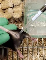
- C57BL/6 male and female mice from Charles River Laboratories
- EpCAM-FITC (CD326) monoclonal antibody (eBioscience, catalog number: 11-5791-80; Clone G8.8)
- Alexa-647 conjugated PECAM antibody (CD31-Alexa 647) (Biolegend, catalog number: 102416; Clone 390)
- Laboratory tissue (Blue roll)
- Metal flat washer (weight 1.66 g) (M8-5/16th inches diameter) (B&Q, UK)
- Absolute ethanol (Sigma-Aldrich, catalog number: 34852-M)
- Pentobarbitone (Pentoject, Animalcare, catalog number: XVD 132)
- Low-melting-point agarose (Sigma-Aldrich, catalog number: A9414)
- Hanks' Balanced Salt Solution (HBSS) (1x) (Life Technologies, catalog number: 14025-050)
- HEPES 1 M (Life Technologies, Gibco, catalog number: 15630080)
- Phosphate Buffered Saline (PBS) (Life Technologies, Gibco, catalog number: 20012068)
- Dulbecco's Modified Eagle Medium (DMEM) (Life Technologies, Gibco, catalog number: 31966-021), also referred to as 'DMEM basal media” in the Recipes
- Penicillin-Streptomycin (10,000 U/ml) (Life Technologies, Gibco, catalog number: 15140122)
- MTT reagent (Thiazolyl Blue Tetrazolium Bromide) (Sigma-Aldrich, catalog number: M2128)
- Dimethyl sulfoxide (DMSO) (Sigma-Aldrich, catalog number: 276855)
- 10% neutral buffered formalin (Sigma-Aldrich, catalog number: HT501128)
- DAPI (Stock concentration 10 mg/ml) (Sigma-Aldrich, catalog number: D9542)
- LIVE/DEAD® Viability/Cytotoxicity Kit (Thermo Fisher Scientific, catalog number: L3224)
- Methanol (Sigma-Aldrich, catalog number: 34860)
- ProLong® Gold Antifade Mountant (Thermo Fisher Scientific, catalog number: P36930)
- Silicon rhodamine far-red fluorophore-conjugated DNA minor groove binder bisbenzimide (SiR-DNA) (tebu-bio Ltd, catalog number: SC007)
- Phenol red-free DMEM with HEPES (Life Technologies, catalog number: 21063029), also referred to as 'Phenol red-free DMEM with HEPES basal media” in the Recipes
- Bovine Serum Albumin (BSA) (Sigma-Aldrich, catalog number: A7030)
- Triton X-100 (Sigma-Aldrich, catalog number: X100)
- (Optional) para-Nitroblebbistatin (Cayman Chemical Company, catalog number: 13891)
- (Optional) Cytochalasin D (Sigma-Aldrich, catalog number: C8273)
- HBSS/HEPES ice cold buffer (see Recipes)
- Agarose solution (see Recipes)
- SF-DMEM (see Recipes)
- Image media (see Recipes)
- MTT working solution (see Recipes)
- 70% ethanol (see Recipes)
- 70% methanol (see Recipes)
Equipment
- Curved dissecting forceps, 10 cm, Serr/C (World Precision Instruments Ltd, catalog number: 15915)
- Fine tip dissecting scissors, 10 cm, straight (World Precision Instruments Ltd, catalog number: 14393)
- Surgical scissors, 14 cm, straight (World Precision Instruments Ltd, catalog number: 14192)
- Spring scissors, 12 cm straight, 12 mm extra-fine blades (World Precision Instruments Ltd, catalog number: 14125)
- Stainless steel blades for vibratome (Campden Instruments LTD, catalog number: 7550-1-SS)
- Automated vibratome (Compresstome® VF-300-0Z; Precisionary Instruments LLC, USA)
- Incubator (Humidified, 37 °C, 5% CO2)
- -20 °C freezer
- GFP filter, excitation 450-490 nm, emission 500-550 nm (for EpCAM-FITC)
- Cy-5 filter, excitation 625-655 nm, emission 665-715 nm (for SiR-DNA and PECAM)
- Plate reader (Tecan; SunriseTM, INSTSUN-1)
- Zeiss Axio Observer inverted widefield microscope, with Lumencor Spectra X LED light source and Hamamatsu Flash 4.0 camera (Zeiss, Germany)
- Zeiss LSM-510 inverted confocal microscope (Zeiss, Germany, model: LSM 510)
- Leica DM2500 widefield microscope (Leica Microsystems, model: Leica DM2500)
Software
- Zen2 acquisition software, blue version (Zeiss, Germany)
- ZEN 2009 (black edition) software (Zeiss, Germany)
- FIJI (ImageJ, version 2.0)
- Icy open source bioimaging analysis software (Version 1.9.8.0; created by the Quantitative Image Analysis Unit at Institut Pasteur, Paris, France)
- Huygens deconvolution software (Scientific Volume Imaging, SVI, Essential version 17.10)
- NIS-Elements (Version 4.50, Nikon Instruments, UK)
- GraphPad Prism version 5
- Microsoft Excel (Microsoft Office 2011 version)
- Microsoft PowerPoint (Microsoft Office 2016 version)
Procedure
文章信息
版权信息
© 2019 The Authors; exclusive licensee Bio-protocol LLC.
如何引用
Akram, K. M., Yates, L. L., Mongey, R., Rothery, S., Gaboriau, D. C. A., Sanderson, J., Hind, M., Griffiths, M. and Dean, C. H. (2019). Time-lapse Imaging of Alveologenesis in Mouse Precision-cut Lung Slices. Bio-protocol 9(20): e3403. DOI: 10.21769/BioProtoc.3403.
分类
细胞生物学 > 细胞成像 >
您对这篇实验方法有问题吗?
在此处发布您的问题,我们将邀请本文作者来回答。同时,我们会将您的问题发布到Bio-protocol Exchange,以便寻求社区成员的帮助。