- EN - English
- CN - 中文
Immunohistochemical Identification of Muscle Fiber Types in Mice Tibialis Anterior Sections
小鼠胫骨前肌切片中肌纤维类型的免疫组化鉴定
发布: 2019年10月20日第9卷第20期 DOI: 10.21769/BioProtoc.3400 浏览次数: 6996
评审: Liang LiuAnonymous reviewer(s)

相关实验方案

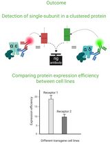
Cluster FLISA——用于比较不同细胞系蛋白表达效率及蛋白亚基聚集状态的方法
Sabrina Brockmöller and Lara Maria Molitor
2025年11月05日 1205 阅读
Abstract
Mammalian skeletal muscle is a metabolically active tissue that is made up of different types of muscle fibers. These myofibers are made up of important contractile proteins that provide force during contraction of the muscle like actin and myosin. Murine myofibers have been classified into 4 types: Type I, Type IIa, Type IIb and Type IIX. Each muscle fiber has been identified with specific type of MyHC expressed, which in turn gives differential contractility to the muscle.
There have been well-known methodologies to identify different myofibers: histochemical myosin ATPase staining which uses the differential ATPase activity between slow and fast fibers, quantification of metabolic enzymes like malate dehydrogenase and lactate dehydrogenase on specific fragments of muscle fibers. The drawback of these techniques is that they cannot differentiate the subtypes of myofibers, for example, Type IIa and Type IIb. They should be used in conjunction with other known histochemical staining techniques. Here, we devise a direct and robust immunohistochemical staining methodology that utilizes the differential expression of MyHC isoforms in different myofibers types, thus efficiently distinguishing the heterogeneity of the muscle fibers. We use antibodies that specifically recognize Type I, Type IIa and Type IIb fibers on serially cut frozen mouse tibialis anterior sections that can be quantified by ImageJ software.
Background
Skeletal muscle is a highly organized tissue composed of several distinct compartments of muscle fibers. Each muscle fiber is a multinucleated single cell, enclosed by sarcolemma, and is predominantly made up of myofibrils (Seeley et al., 2007). Muscle fibers that have low contraction speed are called slow twitch or type I fibers. Slow twitch fibers have high mitochondrial density and express myosin isoform Myh7 (Lompre et al., 1984; Talmadge and Roy, 1993). Type II fibers are divided into type IIa and type IIb fibers. Both of these fibers have high contraction speed, thus generating greater force compared to type I fibers. Type IIa fibers have moderately high contraction speed and are anaerobic in nature. Type IIa fibers also have high density of mitochondria with increased oxidative potential. Type IIa fibers predominantly contain myosin isoform Myh2 in mammals. Type IIb fibers have very high contraction speed and are anaerobic in nature. Type IIb fibers have low mitochondrial density and predominantly contain myosin isoform Myh4. Muscle fibers that are neither Type IIa nor Type IIb fibers are called Type IIx or Type IId, and have high expression in the diaphragm and hence is omitted from this study (Schiaffino and Reggiani, 1994; Westerblad et al., 2010). Mouse tibialis anterior, a hind limb skeletal muscle made of Type I, Type IIa and Type IIb myofibers serves as a suitable muscle to study the robustness of the immunohistochemical staining procedure.
Other methodologies to distinguish the different muscle fiber types are myosin ATPase histochemical staining of frozen muscle sections pre-incubated in alkaline pH showing light staining for Type I fibers and dark staining for Type II fibers. Muscle sections pre-incubated in acidic pH showed dark ATPase staining for Type I fibers, light staining for Type IIa muscle fibers and intermediate staining for Type IIb fibers (Brooke and Kaiser, 1970, Guth and Samaha 1970). Quantitative estimation of mitochondrial enzymes like glycerophosphate oxidase (GP-OX) and succinate dehydrogenase (SDH) have been used to distinguish Type IIa and Type IIb fibers in mouse and rabbit tibialis anterior muscle but gave variable ratios of enzyme activities in mice and rabbit muscles (Reichmann and Pette, 1984). There have been quantification methodologies of specific metabolic enzymes like lactate dehydrogenase and malate dehydrogenase on frozen serial sections of muscles (Hintz et al., 1984). However, these protocols are extensive and may give non-specific signals. The protocol detailed here uses specific antibodies that recognize different Myosin Heavy chain isoforms on serially cut frozen tibialis anterior mid-belly region sections that is readily detected with a suitable secondary antibody. After image acquisition, the number of each fiber type is quantified using ImageJ software.
Materials and Reagents
- High Profile microtome blades 818 (Leica Biosystems, Germany, catalog number: 14035838926)
- Microscope glass slides (Globe Scientific, USA, catalog number: GS-1308)
- Camel hair brushes
- Syringe plunger
- 1.5 ml microfuge tubes (USA Scientific, catalog number: 1615-5500)
- Liquid blocker Pap pen (Sigma Aldrich, catalog number: Z377821)
- Freshly dissected mouse hind limb tibialis anterior muscle
- Myh fast 2A antibody (Developmental Studies Hybridoma Bank, catalog number: 2F7)
- Myh fast 2B antibody (Developmental Studies Hybridoma Bank, Iowa City, IA, catalog number: 10F5)
- Myh slow (type1) antibody (Developmental Studies Hybridoma Bank, Iowa City, IA, catalog number: A4.840)
- Biotinylated sheep anti-mouse IgG antibody (Abcam, catalog number: ab6807)
- Tissue-Tek Optimal cutting temperature (OCT) compound (Sakura Finetek, USA, catalog number: 4583)
- Isopentane (Alfa Aesar, catalog number: 19387, CAS number: 78-78-4)
- Liquid Nitrogen
- Bovine serum albumin (BSA) (Roche, Switzerland, catalog number: 10711454001)
- Normal sheep serum (NSS) (Sigma-Aldrich, catalog number: S22)
- Tween 20 (RPI Corp., USA, catalog number: P20370, CAS number: 9005-64-5)
- 4',6-diamidino-2-phenylindole Dihydrochloride (DAPI) (Invitrogen, catalog number: D1306)
- ProLong Gold Anti-fade solution (Invitrogen, catalog number: P10144)
- Streptavidin Alexa Fluor 488 conjugate (Invitrogen, catalog number: S-11223)
- Sodium phosphate, monobasic
- Sodium phosphate, dibasic
- 37% Formaldehyde (Sigma-Aldrich, catalog number: 252549, CAS number: 50-00-0)
- Milli-Q water
- Blocking mixture (see Recipes)
- Diluent (see Recipes)
- 10% Buffered formalin (see Recipes)
- 0.2 μg/ml DAPI in 1x PBS (see Recipes)
- 1x Phosphate buffered saline (PBS), pH 7.4 (see Recipes)
Equipment
- Rotary Cryostat Microtome (CM1950, Leica Biosystems, Germany)
- Orbital shaker (Boeco, Germany, Ref: BOE 8069200)
- Leica CTR 6500 microscope equipped with Leica DFC 310 FX camera
- Cryostat
Software
- Image J (Fiji, https://imagej.nih.gov/ij/)
Procedure
文章信息
版权信息
© 2019 The Authors; exclusive licensee Bio-protocol LLC.
如何引用
Rao, V. V. and Mohanty, A. (2019). Immunohistochemical Identification of Muscle Fiber Types in Mice Tibialis Anterior Sections. Bio-protocol 9(20): e3400. DOI: 10.21769/BioProtoc.3400.
分类
发育生物学 > 细胞生长和命运决定 > 肌纤维
生物化学 > 蛋白质 > 免疫检测
您对这篇实验方法有问题吗?
在此处发布您的问题,我们将邀请本文作者来回答。同时,我们会将您的问题发布到Bio-protocol Exchange,以便寻求社区成员的帮助。
Share
Bluesky
X
Copy link


