

Isolation and Transcriptomic Profiling of Single Myofibers from Mice
小鼠单根肌纤维的分离和转录组分析

(*contributed equally to this work) 发布: 2019年10月05日第9卷第19期 DOI: 10.21769/BioProtoc.3378 浏览次数: 6857
评审: David PaulClara Lubeseder-MartellatoAmriti Rajender Lulla
参见作者原研究论文
The authors used this protocol in:
Mar 2019
Advertisement


Abstract
Skeletal muscle is composed of different cells and myofiber types, with distinct metabolic and structural features. Generally, transcriptomic analysis of skeletal muscle is performed using whole muscle, resulting in average information as all cells composing the organ contribute to the expression value detected for each gene with the loss of information about the distinctive features of each specific myofiber type. Since myofibers are the smallest complete contractile system of skeletal muscle influencing its contraction velocity and metabolism, it would be beneficial to have fiber-specific information about gene expression. Here, we describe a protocol for the isolation and the transcriptomic analysis of single individual myofibers. The protocol was set up using single myofibers isolated from soleus and Extensor Digitorum Longus (EDL) muscles, but it can be applied to all skeletal muscles. Briefly, muscles are enzymatically dissociated and individually collected. Long RNAs (> 200 nt) and short RNAs (< 200 nt) are separately purified from each myofiber and used to produce libraries for microarray or sequencing analysis. Through this approach, myofiber-specific transcriptional profiles can be produced, free from transcripts from other non-contractile cell types, in order to identify mRNA-miRNA-lncRNA regulatory networks specific for each myofiber type.
Keywords: Single myofiber (单根肌纤维)Background
Skeletal muscle is a complex organ and heterogenous tissue. It is composed by different cell types such as those from blood, vessels, nerves, connective tissue, and myofibers, that are the smallest complete contractile system of skeletal muscle influencing both its mechanical performance and metabolism. In addition, myofibers are classified into different types. In mouse, type I myofibers display slow-twitch contraction and oxidative metabolism, type IIA and IIX myofibers fast-twitch contraction and oxidative metabolism, and type IIB myofibers fast-twitch contraction and glycolytic metabolism (Schiaffino and Reggiani, 2011). The preferential metabolism of each muscle is defined by the different proportion of myofiber types. Soleus muscle is mainly composed by type I, type IIA, and type 2X oxidative myofibers, and preferentially consumes lipids, whereas EDL muscle by type IIB glycolytic myofibers, and primarily uses carbohydrates as energy substrate (Augusto et al., 2004). To investigate differences in muscle metabolism and contraction at transcriptomic level, previous expression profiles were obtained using soleus and EDL whole muscles (Campbell et al., 2001; Wu et al., 2003). Results described in these works are influenced by the different contributions of diverse cells composing whole muscle (Figure 1A) and lose the transcriptional specificity of myofiber types (Figure 1B). We demonstrated the feasibility of scaling down the transcriptomic analysis of skeletal muscle at single isolated myofiber level for mRNA, long non-coding RNA (lncRNA) and microRNA (miRNA) populations (Figure 1C). This approach allowed the identification of expression profiles, free from non-myofiber cells (Chemello et al., 2011), and allowed the generation of mRNA-miRNA-lncRNA regulatory networks specific for each myofiber type (Alessio et al., 2019; Chemello et al., 2019). Moreover, it is easier to characterize perturbations that exclusively affect myofibers (Chemello et al., 2015; Mammucari et al., 2015), and to improve the description and comprehension of muscle atrophy (Alessio et al., 2019). In fact, muscle atrophy induces significant systemic metabolic modifications (Celegato et al., 2006) also in a myofiber-specific manner (Wang and Pessin, 2013).
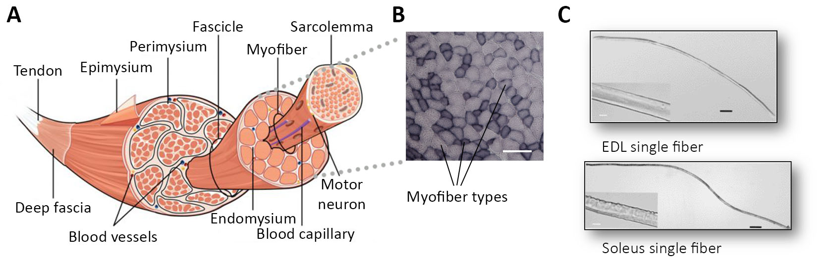
Figure 1. Complexity of skeletal muscle. A. Structure of the skeletal muscle with the different tissues and cells that make up its structure. Image modified from (Biga et al., 2019). B. Succinate Dehydrogenase Staining (SDH) shows different myofiber types in a transversal muscle cross-section. Scale bar is for 100 µm. C. Images of single isolated myofibers from EDL and soleus muscles. Scale bar is for 250 µm. From each single myofibers is possible to profile the expression of long and short RNAs.
Materials and Reagents
- 200 µl tips
- DNA LoBind tubes (Eppendorf, catalog number: 0030108051 or Serstedt, catalog number: 72.706.700)
- Spin-X centrifuge tubes (Corning-Costar, catalog number: CLS8160-96EA)
- RNase/DNase free Eppendorf microcentrifuge tube
- 0.2 ml PCR tube
- Hybridization gasket slides, 8 microarrays/slide (Agilent, catalog number: G2534-60014)
- Cell culture plates: 24-well and 6-well plates
- Plastic Pasteur pipettes
- Slide-staining dishes
- Adult wild-type mice, weight: 33-35 g
Note: We used CD1 mouse strain. - High-glucose Dulbecco's modified Eagle medium (DMEM) (Millipore Sigma, catalog number: D5796)
- 2,3-Butanedione monoxime (BDM) (Sigma-Aldrich, catalog number: B0753)
- Agencourt AMPure XP beads (Beckman Coulter, catalog number: A63881)
- Collagenase from Clostridium histolyticum, type I (Millipore Sigma, catalog number: C0130)
- Fetal bovine serum (FBS) (Millipore Sigma, catalog number: F2442)
- Phosphate-buffered saline (PBS) (Thermo Fisher Scientific, catalog number: 20012019)
- TRIzol Reagent (Thermo Fisher Scientific, catalog number: 15596026)
- Nuclease-free water (Thermo Fisher Scientific, catalog number: 10977035)
- Nuclease-free water (Ambion, catalog number: AM9937)
- Chloroform (PanReac AppliChem, catalog number: 121252)
- 70% v/v ethanol (dilute ethanol in nuclease-free water)
- 75% v/v ethanol (dilute ethanol in nuclease-free water)
- 80% v/v ethanol (dilute ethanol in nuclease-free water)
- Absolute ethanol
- miRNeasy kit (Qiagen, catalog number: 217004)
- RNeasy micro kit (Qiagen, catalog number: 74004)
- Complete Whole Transcriptome Amplification Kit (Millipore Sigma, catalog number: WTA2)
- GenElute PCR Clean-Up Kit (Millipore Sigma, catalog number: NA1020)
- SureTag DNA Labeling Kit (Agilent, catalog number: 5190-3400)
- Gene Expression Hybridization Kit (Agilent, catalog number: 5190-0404)
- Gene Expression Wash Buffer Kit (Agilent, catalog number: 5188-5327)
- SurePrint G3 Mouse GE 8x60K Microarray Kit (Agilent, catalog number: G4852A)
- Poly(A) Tailing Kit (Thermo Fisher Scientific, catalog number: AM1350)
- Sodium Acetate 3 M (pH 5.5) (Thermo Fisher Scientific, catalog number: AM9740)
- SuperScript II Reverse Transcriptase (Thermo Fisher Scientific, catalog number: 18064014)
- Oligo-dT-Ion P1 adapter primer (5′-CCTCTCTATGGGCAGTCGGTGATCCTCAGC[dT]20VN-3′)
- SMART primer (5′-CACACACAATTAACCCTCACTAAAggg-3′)
- Ion Xpress Plus gDNA Library (Thermo Fisher Scientific, catalog number: 4471269)
- PGM HI-Q OT2 kit (Thermo Fisher Scientific, catalog number: A27739)
- Platinum Taq DNA Polymerase High Fidelity (Thermo Fisher Scientific, catalog number: 11304011)
- E-Gel SizeSelect Gels (Thermo Fisher Scientific, catalog number: G661012)
- DNA ladder 50 bp (Thermo Fisher Scientific, catalog number: 10416014)
- High Sensitivity DNA kit (Agilent, catalog number: 5067-4626)
- Ion OneTouch Template Kit (Thermo Fisher Scientific, catalog number: A29900)
- 1x Low TE buffer (see Recipes)
Equipment
- Micropipette (P10, P20)
- -80 °C freezer
- -20 °C freezer
- Forceps and scissors for microdissection
- 37 °C incubator
- Stereo-microscope
- Refrigerated microcentrifuge
- SpeedVac concentrator
- Thermocycler
- Nanodrop spectrophotometer
- Heat block
- Centrifuge
- Agilent SureHyb chamber (Agilent, catalog number: G2534A)
- Hybridization oven (Agilent, model: G2545A)
- Hybridization oven rotator (Agilent, model: G2530-60029)
- Agilent scanner system (Agilent, model: G2505C)
- DynaMag (Thermo Fisher Scientific, catalog number: 12321D)
- Ion Torrent, or Ion S5 System (Thermo Fisher Scientific)
- E-GelTM Electrophoresis Device (Thermo Fisher Scientific)
- Agilent 2100 Bioanalyzer (Agilent)
- Ion OneTouch Instrument (Thermo Fisher Scientific)
- Ion PGM sequencing instrument (Thermo Fisher Scientific)
Software
- Bioanalyzer software (Agilent, https://www.agilent.com/en/product/automated-electrophoresis/bioanalyzer-systems/bioanalyzer-instrument/2100-bioanalyzer-laptop-228251 Company or Developer/Provider/Supplier, web address)
- Feature Extraction Software (Agilent, https://www.agilent.com/en/product/mirna-microarray-platform/mirna-microarray-software/feature-extraction-software-228496)
- R statistical software (https://www.r-project.org/)
- MultiExperiment Viewer (https://sourceforge.net/projects/mev-tm4/)
- miRDeep software (https://sourceforge.net/projects/mirdeepstar/)
Procedure
文章信息
版权信息
© 2019 The Authors; exclusive licensee Bio-protocol LLC.
如何引用
Chemello, F., Alessio, E., Buson, L., Pacchioni, B., Millino, C., Lanfranchi, G. and Cagnin, S. (2019). Isolation and Transcriptomic Profiling of Single Myofibers from Mice. Bio-protocol 9(19): e3378. DOI: 10.21769/BioProtoc.3378.
分类
分子生物学 > RNA > 转录
分子生物学 > RNA > miRNA与mRNA 的相互作用
您对这篇实验方法有问题吗?
在此处发布您的问题,我们将邀请本文作者来回答。同时,我们会将您的问题发布到Bio-protocol Exchange,以便寻求社区成员的帮助。