

QUEEN-based Spatiotemporal ATP Imaging in Budding and Fission Yeast
发芽酵母和裂殖酵母中基于QUEEN的时空ATP成像
发布: 2019年08月05日第9卷第15期 DOI: 10.21769/BioProtoc.3320 浏览次数: 7128
评审: Samantha E. R. DundonUdita UpadhyayGunjan Mehta
参见作者原研究论文
The authors used this protocol in:
Apr 2019
Advertisement


Abstract
Yeasts have provided an exceptional model for studying metabolism and bioenergetics in eukaryotic cells. Among numerous metabolites, adenosine triphosphate (ATP) is a major metabolite that is essential for all living organisms. Therefore, a clearer understanding of ATP dynamics in living yeast cells is important for deciphering cellular energy metabolism. However, none of the methods currently available to measure ATP, including biochemical analyses and ATP indicators, have been suitable for close examinations of ATP concentrations in yeast cells at the single cell level. Using the recently developed ATP biosensor QUEEN, which is suitable for yeasts and bacteria, a protocol was described herein to visualize ATP concentrations in living budding and fission yeast cells. This simple method enables the easy and reliable examination of ATP dynamics in various yeast mutants, thereby providing novel molecular insights into cellular energy metabolism.
Keywords: ATP (ATP)Background
Adenosine triphosphate (ATP) is one of the major and indispensable metabolites for all living organisms. In addition to being useful “energy currency” for various cellular processes, ATP serves as an intracellular and extracellular signaling molecule as well as a phosphate donor for protein phosphorylation (Lehninger et al., 2010). Recent studies suggested that ATP itself solubilizes protein as a “biological hydrotrope” (Patel et al., 2017) and influences the aggregation state of intracellular proteins (Pu et al., 2019; Sridharan et al., 2019). Due to the obvious importance of ATP and its wide involvement in many cellular events, the spatial distributions and temporal dynamics of ATP in living organisms need to be clarified in order to elucidate energy metabolism and signal transductions at the molecular level. To date, a number of methods have been developed to measure ATP concentrations in vitro. However, these biochemical methods have only a limited ability to examine ATP dynamics in vivo because of their insufficient time resolution relative to the rapid turnover of ATP (less than one minute) (Mortensen et al., 2011; Takaine et al., 2019). Moreover, although these biochemical analyses detect the average ATP level of a cell population, they are incapable of showing the intracellular or intratissue distribution of ATP.
To overcome these difficulties, several types of ATP indicators have been developed for the direct visualization of ATP in living organisms (Dong and Zhao, 2016). Among them, genetically encodable ATP indicators are advantageous because their introduction into a cell population is non-invasive and highly specific. A series of fluorescence resonance energy transfer (FRET)-based genetically encoded ATP indicators, named “the ATeam”, was developed for the first time a decade ago by the pioneering work of Imamura’s group (Imamura et al., 2009). The ATeam comprises the ATP-binding domain of the ε subunit of bacterial FoF1 ATP synthase and flanking cyan and yellow fluorescent proteins (FPs), and is now widely and successfully used to monitor intracellular ATP concentrations in various types of cells ranging from plants to mammals. However, the ATeam composed of two FPs may be susceptible to proteolysis and, thus, yield immature sensor molecules because of differences in the maturation times of the two FPs. These features manifest when the ATeam is expressed in fast-growing microorganisms, in which protein turnover is very rapid, such as yeasts or bacteria, and become problematic because the presence of a dysfunctional sensor inevitably affects the FRET signal. To circumvent the drawbacks associated with ATeam, the second-generation ATP indicator “QUEEN” (quantitative evaluator of cellular energy) was recently developed by Imamura’s group (Yaginuma et al., 2014). QUEEN is a dual-excitation ratiometric ATP biosensor composed of a single FP and, thus, is more resistant to degradation and matures more rapidly than ATeam, yielding the reliable quantification of ATP, even in actively proliferating bacterial cells.
Yeasts have provided an exceptional model for studying metabolism and bioenergetics in eukaryotic cells. Yeast carbon metabolism has long been studied because of its industrial importance for producing many valuable chemicals (yeast metabolites) (Borodina and Nielsen, 2014). Energy metabolism in yeasts also provides a tractable model for studying energy metabolism in tumor cells because they are similar in that they synthesize the majority of ATP through glycolysis and not oxidative phosphorylation, even in the presence of oxygen, and this is known as “aerobic glycolysis” or “the Warburg effect” (Diaz-Ruiz et al., 2011). Therefore, a close examination of ATP dynamics in living yeast cells has been of great significance, but waits to be embarked due to the absence of an appropriate ATP biosensor.
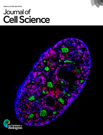
In a recent study, QUEEN was applied to both the budding yeast Saccharomyces cerevisiae and fission yeast Schizosaccharomyces pombe for the first time and intracellular ATP levels and its dynamicity were visualized in wild-type cells (Takaine et al., 2019). The use of QUEEN enables the easy and reliable examination of ATP dynamics in living yeast cells, providing novel insights into cellular energy metabolism. The protocols used to observe and analyze the QUEEN fluorescence signal in yeast cells have been described herein. The constitutive and strong expression of QUEEN did not induce any noticeable defects in cell growth, meiosis, or sporulation. Thus, ATP concentrations in various yeast mutants under a number of conditions may be observed, as well as ATP in wild-type cells, using the same method. Moreover, this method employs a conventional wide-field fluorescence microscope and free image processing software for image calculations, and, thus, is accessible to a broad range of researchers.
Materials and Reagents
- 35-mm glass-bottomed dish (IWAKI, catalog number: 3971-035, No. 1.5 thickness)
- Cell culture dish, sterilized (SANSEI MEDICAL CO., LTD., catalog number: 01-013)
- Pipette tips (Watson® Bio Lab, catalog numbers: 123R-755YS [200 µl], 123R-757CS [1,000 µl])
- Sterile culture tube (16 ml, polypropylene) (EVERGREEN, catalog number: 222-2393-080)
- Toothpicks (6.5-18 cm) (any brand)
Note: Sterilize before use. Reusable until they break. - Budding yeast cells expressing QUEEN-2m:
e.g., MTY3261 (MATa his3∆1::3×pRS303-PTEF1-QUEEN-2m-TCYC1 leu2∆0 ura3∆0) from Takaine et al., 2019. This strain was derived from a slightly modified BY4741 parental strain (MATa his3∆1 leu2∆0 ura3∆0). - Fission yeast cells expressing QUEEN-2m:
e.g., KSP3769 (h- leu1-32::Ptif51-QUEEN-2m::leu1+) from Ito et al., 2019.
Note: Both yeast strains expressing QUEEN-2m and plasmids for expressing QUEEN-2m in budding yeast are available from the Yeast Genetic Resource Centre Japan (YGRC, http://yeast.nig.ac.jp/yeast/). - Concanavalin A (Sigma-Aldrich, catalog number: C-7275)
- Soybean lectin (Sigma-Aldrich, catalog number: L-1395)
- Yeast nitrogen base w/o amino acids (YNB) (Invitrogen, catalog number: Q300-09)
- Yeast extract (BD, catalog number: 212750)
- Bacto-peptone (BD, catalog number: 211677)
- D(+)-Glucose (FUJIFILM Wako, catalog number: 049-31165)
- 2-Deoxy-D-glucose (2DG) (FUJIFILM Wako, catalog number: 046-06483)
Note: 2DG is a potent inhibitor of glycolysis and has been used to deplete ATP in yeast cells (Serrano, 1977; Xu and Bretscher, 2014; Takaine et al., 2019). - Agar (FUJIFILM Wako, catalog number: 010-08725)
- Potassium hydrogen phthalate (FUJIFILM Wako, catalog number: 163-03822)
- Na2HPO4·12H2O (Kishida Chemical Co., Ltd., catalog number: 000-72535)
- Ethanol (Kishida Chemical Co., Ltd., catalog number: 140-28553)
- NH4Cl (FUJIFILM Wako, catalog number: 017-02995)
- Uracil (FUJIFILM Wako, catalog number: 212-00062)
- Myo-inositol (FUJIFILM Wako, catalog number: 096-00285)
- L-Glutamic acid (FUJIFILM Wako, catalog number: 072-00501)
- Adenine sulfate (FUJIFILM Wako, catalog number: 018-19613)
- L-Leucine (PEPTIDE INSTITUTE INC., catalog number: 2713)
- L-Histidine (FUJIFILM Wako, catalog number: 082-00683)
- L-Lysine·HCl (PEPTIDE INSTITUTE INC., catalog number: 2714)
- L-Methionine (PEPTIDE INSTITUTE INC., catalog number: 2715)
- L-Phenylalanine (PEPTIDE INSTITUTE INC., catalog number: 2717)
- L-Serine (PEPTIDE INSTITUTE INC., catalog number: 2719)
- L-Threonine (PEPTIDE INSTITUTE INC., catalog number: 2720)
- MgCl2·6H2O (FUJIFILM Wako, catalog number: 132-00175)
- CaCl2·2H2O (FUJIFILM Wako, catalog number: 033-25035)
- KCl (FUJIFILM Wako, catalog number: 163-03545)
- Na2SO4 (FUJIFILM Wako, catalog number: 195-03341)
- H3BO3 (FUJIFILM Wako, catalog number: 021-02195)
- MnSO4·5H2O (FUJIFILM Wako, catalog number: 139-00825)
- ZnSO4·7H2O (FUJIFILM Wako, catalog number: 268-00405)
- FeCl3·6H2O (FUJIFILM Wako, catalog number: 095-00875)
- Na2MoO4·2H2O (FUJIFILM Wako, catalog number: 198-02471)
- KI (FUJIFILM Wako, catalog number: 166-03971)
- CuSO4·5H2O (FUJIFILM Wako, catalog number: 031-04411)
- Citric acid (FUJIFILM Wako, catalog number: 038-05521)
- Nicotinic acid (FUJIFILM Wako, catalog number: 142-01232)
- Biotin (TOKYO CHEMICAL INDUSTRY CO., LTD., catalog number: B0463)
- Sodium pantothenate (FUJIFILM Wako, catalog number: 198-05651)
- 100x Amino acids mix (see Recipes)
- 100x L-Leucine stock (see Recipes)
- 50x Uracil stock (see Recipes)
- SC-His liquid medium (see Recipes)
- YPD agar plate (see Recipes)
- YELA agar plate (see Recipes)
- 100x 5 Low-supplements (see Recipes)
- 50x Salt stock (see Recipes)
- 1,000x Vitamins (see Recipes)
- 10,000x Minerals (see Recipes)
- EMM liquid medium (see Recipes)
- 2-Deoxy-D-glucose (2DG) medium (see Recipes)
- Glass-bottomed dish coated with concanavalin A (see Recipes)
- Glass-bottomed dish coated with soybean lectin (see Recipes)
Equipment
- Inverted fluorescent microscope (Nikon, Eclipse Ti-E) equipped with:
- A 100x objective lens (Nikon, Apo TIRF 100x Oil DIC N2/NA 1.49)
- A high-pressure mercury lamp (Nikon, Intensilight C-HGFIE 130W)
- An electron multiplying charge-coupled device camera (Andor-Oxford Instruments, iXon3 DU897E-CS0-#BV80)
- A FITC filter set (Nikon, Ex465-495/DM505/BA515-555)
- A custom-made Ex409 filter set (Semrock, Ex393-425/DM506/BA516-556)
- Neutral density (ND) filters (Nikon, ND4 [MBN21804] and ND8 [MBN21808])
- Bench-top aspirator (INTEGRA, VACUSIP)
- Inverted routine microscope (Nikon, Eclipse Ts2) equipped with a 40× objective lens (Nikon, CFI Achromat LWD ADL 40XF/NA 0.55, catalog number: MRP46402)
- Autoclave (TOMY SEIKO CO., LTD, LBS-325)
- Pure water system (Merck Millipore, Milli-Q® Integral 15)
- Erlenmeyer flasks (50 ml) (HARIO, catalog number: 82-0144)
- Silicone stopper (for a 50-ml flask) (Shin-Etsu Polymer, SILICOSEN T22)
- Incubator (TAITEC, INCUBATE BOX M-200F)
- Culture rotator (TAITEC, RT-50)
- Rotary shaker (NISSIN, NX-20)
- Pipettes (BM EQUIPMENT Co., LTD, PipetPAL®, model: PAL-200 [20-200 µl], PAL-1000 [100-1,000 µl])
Software
- NIS-Element AR ver. 4.30.01 (Nikon, https://www.microscope.healthcare.nikon.com/ja_JP/)
- Fiji ver. 2.0.0-rc-69/1.52n (Fiji contributors [Schindelin et al., 2012], http://fiji.sc/#)
- Microsoft® Excel for Mac 2011 ver. 14.7.3 (Microsoft, https://products.office.com/ja-jp/excel)
- KaleidaGraph ver. 4.5.1 (HULINKS, https://www.hulinks.co.jp/software/stat_graph/kaleida)
Procedure
文章信息
版权信息
© 2019 The Authors; exclusive licensee Bio-protocol LLC.
如何引用
Takaine, M. (2019). QUEEN-based Spatiotemporal ATP Imaging in Budding and Fission Yeast. Bio-protocol 9(15): e3320. DOI: 10.21769/BioProtoc.3320.
分类
细胞生物学 > 细胞成像 > 荧光
微生物学 > 体内实验模型 > 真菌
您对这篇实验方法有问题吗?
在此处发布您的问题,我们将邀请本文作者来回答。同时,我们会将您的问题发布到Bio-protocol Exchange,以便寻求社区成员的帮助。