- EN - English
- CN - 中文
Isolation of Powdery Mildew Haustoria from Infected Barley
感染大麦中白粉病吸器的分离提取
发布: 2019年07月20日第9卷第14期 DOI: 10.21769/BioProtoc.3299 浏览次数: 7828
评审: Amey RedkarThomas TorodeHailong Guo

相关实验方案

基于 Emu 的可重复计算流程:在高性能计算集群上实现高保真土壤—植物微生物组解析
Henrique M. Dias [...] Christopher Graham
2026年01月20日 386 阅读
Abstract
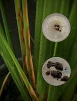
Blumeria graminis is a fungus that causes powdery mildews on grasses, such as barley. Investigations of this pathogen present many challenges due to its obligate biotrophic nature. This means that the fungus can only grow in the presence of a living host plant. B. graminis forms epiphytic mycelia on the plant surface and feeding organs (haustoria) inside the epidermal cells of the host plant. Therefore, it is difficult to separate the fungus from plant tissues. This protocol shows how to obtain different fungal structures from powdery mildew infected barley leaves. The epiphytic mycelia including conidia and conidiophores can be separated after immersing the infected leaves into 5% cellulose acetate dissolved in acetone, and peeling off the cellulose acetate membrane. Then, the haustoria are isolated from dissected epidermis after cellulase degradation of plant cell walls. The isolated haustoria remain intact with few plant impurities. The haustoria may be visualized by epifluorescence microscopy after staining with the chitin-specific dye WGA-Alexa Fluor 488. Finally, dissected material can be either processed immediately or kept at -80 °C for long-term storage for studies on gene expression and protein identification, for example by mass spectrometry.
Keywords: Powdery mildew (白粉病)Background
Powdery mildew of cereals, caused by Blumeria graminis, leads to significant yield loss (Spanu et al., 2010). The barley powdery mildew fungus B. graminis f. sp. hordei, is economically important, and one of the best studied powdery mildews (Both et al., 2005a; Bindschedler et al., 2009). This fungus is an obligate biotroph, which means it can only infect and propagate on living barley plants (Pedersen et al., 2012). Its asexual life cycle involves an intimate relationship with the host plant. Airborne conidia germinate on the host producing first a primary and then a secondary germ tube. The secondary germ tube develops an appressorium, from which a hyphal peg penetrates into an epidermal cell. Inside the cell, a penetration peg enlarges and forms a multidigitate feeding structure: the haustorium. This is surrounded by a plant-derived extrahaustorial membrane (Both et al., 2005b; Bindschedler et al., 2016).
Several studies of B. graminis f. sp. hordei have characterized gene expression profiles and proteomics of this fungus (Bindschedler et al., 2011; Pennington et al., 2016). However, the obligate biotrophic nature of B. graminis poses exceptional challenges to the investigations of this fungus, especially the studies of haustoria. The intracellular haustorium and the extrahaustorial membrane constitute a special compartment, which is functionally essential for the interaction with the host (Wang et al., 2009; Pliego et al., 2013). Investigating haustoria is critical for further understanding the mechanism of nutrient uptake into the pathogen as well as plant-pathogen recognition. Therefore, obtaining purified haustoria from infected plants has the potential of contributing significantly to these studies.
Previously, the procedure for isolating haustoria from obligate biotrophs employed affinity chromatography on rust fungi (Catanzariti et al., 2006; Garnica et al., 2014). During the 1990s, several centrifugation-based methods were introduced to isolate powdery mildew haustoria from pea plants (Mackie et al., 1991; Testut et al., 1999). This method was then applied to powdery mildews of other plants, including Arabidopsis (Wang et al., 2009; Micali et al., 2011). More recently, a filtration and gradient centrifugation-based method was described to isolate powdery mildew haustoria from barley (Godfrey et al., 2009). The hallmark of these published methods is that they are relatively laborious and require several different kinds of isolation buffers, different sizes of steel meshes and multiple centrifugation steps. Besides, the isolation procedure was required to be performed on ice. In this protocol, we developed a simpler way to isolate B. graminis haustoria from infected barley leaves. Releasing of haustoria from plant cells is achieved by enzymatically degrading the epidermal cell walls, and the purification of haustoria by filtration through a nylon mesh. This method requires digestion of the host plant cell walls, a single filtration and one centrifugation step.
Materials and Reagents
- Pipette tips
- Dedicated enclosed transparent container
- Razor blade
- Parafilm
- Pasteur pipettes
- 15 ml/50 ml polypropylene tubes
- Liquid nitrogen container
- Microscope glass slide
- Cover glass
- Barley (Hordeum vulgare) cv. Golden Promise seedlings, barley seeds were sown in John Innes 1 compost mixed with vermiculite (4:1) in 12 x 12 cm pots
- Liquid nitrogen
- Cellulose acetate
- Anhydrous acetone
- MES hydrate powder
- HCl
- WGA, Alexa Fluor 488 conjugate (Thermo Fisher Scientific, catalog number: W11261)
Note: This dye exhibits the bright, green fluorescence of the Alexa Fluor® 488 dye (excitation/emission maxima ~495/519 nm). - Cellulase Onozuka R-10 (Duchefa Biochemie, catalog number: C8001)
Note: “Cellulase Onozuka R-10” from Trichoderma viride.1 unit (U) of Cellulase will release 1.0 μmole of glucose from carboxymethyl cellulose. - NaCl
- KCl
- Na2HPO4
- KH2PO4
- PBS buffer (see Recipe 1)
Equipment
- Forceps
- Pipettes
- Scissor
- Tweezers
- 70 μm nylon mesh sieve
- Incubator
- Centrifuge
- GFP filter
- Epifluorescence microscope
- -80 °C freezer
Procedure
文章信息
版权信息
© 2019 The Authors; exclusive licensee Bio-protocol LLC.
如何引用
Li, L., Collier, B. and Spanu, P. D. (2019). Isolation of Powdery Mildew Haustoria from Infected Barley. Bio-protocol 9(14): e3299. DOI: 10.21769/BioProtoc.3299.
分类
植物科学 > 植物免疫 > 宿主-细菌相互作用
细胞生物学 > 细胞分离和培养 > 细胞分离
微生物学 > 微生物-宿主相互作用 > 真菌
您对这篇实验方法有问题吗?
在此处发布您的问题,我们将邀请本文作者来回答。同时,我们会将您的问题发布到Bio-protocol Exchange,以便寻求社区成员的帮助。
Share
Bluesky
X
Copy link


