

Gentamicin Protection Assay to Determine the Number of Intracellular Bacteria during Infection of Human TC7 Intestinal Epithelial Cells by Shigella flexneri
庆大霉素保护试验确定福氏志贺菌侵染人TC7肠上皮细胞的胞内寄生数目


发布: 2019年07月05日第9卷第13期 DOI: 10.21769/BioProtoc.3292 浏览次数: 9795
评审: Meenal SinhaRAVIKANTH NANDURIAnonymous reviewer(s)
参见作者原研究论文
The authors used this protocol in:
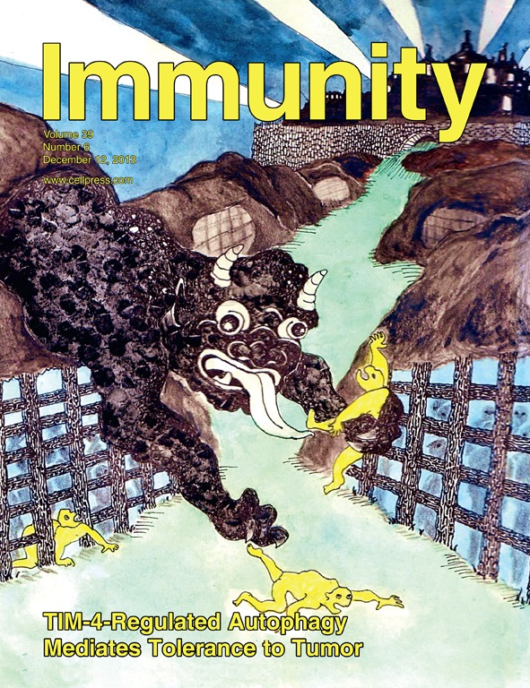
Dec 2013
Advertisement


Abstract
Shigella flexneri is an intracellular bacterial pathogen that gains access to the gut epithelium using a specialized Type III Secretion System (T3SS). Various determinants mediating this invasive infection have been experimentally verified using the classical gentamicin protection assay presented here. In this assay epithelial cell lines are infected by bacteria in vitro and the extracellular bacteria are killed by gentamicin. The internalized bacteria, which are protected from the bactericidal action of gentamicin, are recovered by lysing the epithelial cells and enumerated by determining the colonies formed on solid medium. Various techniques based on light microscopy, such as immunofluorescence and bacteria expressing fluorescent proteins, are also used for studying intracellular bacteria. However, these techniques are not only labor intensive and require sophisticated equipment, but mostly are also not quantitative. Despite being an easy quantitative method to study invasiveness of bacteria, the gentamicin protection assay cannot distinguish between the survival and multiplication of the internalized bacteria over longer incubation periods. To alleviate the complications created by multiplication and dissemination of internalized bacteria, complementary assays like plaque formation assays are required. This protocol presents an easy and cost-effective method to determine the invasiveness and the capacity to establish an infection of Shigella under different conditions.
Keywords: Shigella (志贺氏菌)Background
Shigella infects about 160 million people leading to about 600,000 deaths every year (Reference 8). The clinical manifestations of Shigella infection, or shigellosis, arise only after the bacterium enters the epithelium, where it multiplies and spreads to adjacent cells causing cell death and tissue necrosis. Thus, the entry into the epithelial cells is a critical step in the infectious life cycle of Shigella (Ashida et al., 2015). Most of the determinants mediating this step have been ascribed to a large virulence plasmid that encodes a T3SS responsible for injecting bacterial proteins into the host cell via a needle-like projection (Puhar and Sansonetti, 2014).
The gentamicin protection assay is a classical method that is used to assess the invasiveness of Shigella and has led to the identification of a number of mediators of invasion by mutational analysis and comparisons with the wild-type bacteria. In a typical experiment, the bacteria are allowed to infect the intestinal cells in a synchronized manner followed by removal of external bacteria by gentamicin treatment. The invasiveness is assessed by determining the number of surviving bacteria (protected from gentamicin being intracellular) from the lysates of infected cells. A lower number of surviving bacteria with respect to the wild-type indicates a defect in invasion or intracellular survival. If short infection times are used after the gentamicin treatment (typically 1h), the gentamicin protection assay allows to compare the bacterial capacity to enter host cells only. However, if longer infection times are used after gentamicin treatment (2 h or more), the assay will also reflect the bacterial capacity to survive and multiply within the target cell besides the capacity to invade. Hence, if the gentamicin protection assay is employed to study the capacity to survive and multiply by allowing long infection times, these experiments should always be complemented with tests carried out at short infection times to ensure correct interpretation of the results. The gentamicin protection assay, however, is not suitable to assess the extent of intercellular spread of bacteria throughout the monolayer, which is conveniently determined by a plaque formation assay. Although the invasiveness of bacterial pathogens can also be studied by fluorescence microscopy-based techniques such as immunofluorescence and FACS; it requires the use of costly reagents like labeled probes, special reporter strains and sophisticated equipment. The gentamicin protection assay is not only relatively easy to perform and cheap, it is fairly rapid and less labor intensive making it amenable to higher throughput.
The protocol presented here reprises the classical gentamicin protection assay, which has been optimized for use with Shigella flexneri and TC7 intestinal epithelial cells but can be modified to be used with other intracellular pathogens and other host cell types.
Materials and Reagents
- Conical flask (Duran, catalog number: 2121628)
- Microfuge tubes (Eppendorf, Safe-Lock tubes, catalog numbers: 0030120086, 0030120094)
- Centrifuge tubes (Sarstedt, catalog numbers: 62.554.502, 62.547.254)
- Culture tubes (TPP, catalog number: 91016)
- Pipette tips (VWR, catalog numbers: 89041-404, 89041-412, 89041-400)
- Serological pipettes (VWR, catalog numbers: 612-3702, 612-3700, 612-3698)
- 6-well tissue culture test plates (TPP, catalog number: 92406)
- TC7 cells (human cell line, a derivative of Caco-2 colon adenocarcinoma cells) (Chantret et al., 1994)
- Shigella flexneri M90T (Sansonetti et al., 1982)
- Dulbecco's Modified Eagle Medium (DMEM) (Gibco, catalog number: 21885-025)
- Penicillin-Streptomycin solution (10,000 U/ml) (Gibco, catalog number: 15140122)
- Minimum Essential Medium Non-Essential Amino Acids solution (100x) (Gibco, catalog number: 11140-035)
- Dulbecco’s Phosphate Buffered Saline (PBS) solution (Gibco, catalog number: 14190-144)
- Fetal Bovine Serum (Gibco, catalog number: 10500056)
- HEPES 1 M solution (Gibco, catalog number: 15630-080)
- Trypsin-EDTA (0.05%), phenol red (Gibco, catalog number: 25300-054)
- Trypan Blue Stain (0.4%) (Thermo Scientific, catalog number: T10282)
- Agarose (VWR Life Science, catalog number: 35-1020)
- Tryptic Soy Broth (TSB) ready to use powder (Merck, catalog number: 105459)
- Tryptic Soy Agar (TSA) ready to use powder (Merck, catalog number: 105458)
- Ethanol (VWR chemicals, catalog number: 20821.558)
- Congo red (Sigma, catalog number: C6277)
- Sodium deoxycholate (Sigma, catalog number: 30970)
- Gentamicin sulfate (Sigma, catalog number: G1264)
- Sterile disposable petriplates (Sigma, catalog number: P5606-400EA)
- Congo red solution (see Recipes)
- Growth medium (see Recipes)
- Infection buffer (see Recipes)
- Sodium Deoxycholate solution (see Recipes)
- Gentamicin solution (see Recipes)
Equipment
- Biosafety cabinet (Thermo Scientific, HERAsafe KS 18)
- Spectrophotometer (Amersham, Utrospec 2100 pro)
- Pipette controller (VWR, Accurpette)
- CO2 Incubator (Thermo Scientific, Heracell VIOS 160i)
- Benchtop centrifuge (VWR, Micro Star 17R)
- Centrifuge (Eppendorf, 5810R with Rotor A-4-81 for plates)
- Water bath (Grant, JBA 12)
- Pipettes (Eppendorf, Research plus)
- Inverted microscope (Motic, AE2000 Binocular)
- Cell counter (Thermo Scientific, Countess II FL)
- Cell counter slides (Thermo Scientific, Countess Cell Counting Chamber Slides, C10228)
- Orbital shaker (Edmund Bühler, Swip SM25)
- Vacuum pump (VWR, Mini diaphragm vacuum pump VP 86)
Software
- Prism 7 (GraphPad, https://www.graphpad.com)
Procedure
文章信息
版权信息
© 2019 The Authors; exclusive licensee Bio-protocol LLC.
如何引用
Sharma, A. and Puhar, A. (2019). Gentamicin Protection Assay to Determine the Number of Intracellular Bacteria during Infection of Human TC7 Intestinal Epithelial Cells by Shigella flexneri. Bio-protocol 9(13): e3292. DOI: 10.21769/BioProtoc.3292.
分类
微生物学 > 微生物-宿主相互作用 > 细菌
免疫学 > 宿主防御 > 人
细胞生物学 > 基于细胞的分析方法 > 细菌感染
您对这篇实验方法有问题吗?
在此处发布您的问题,我们将邀请本文作者来回答。同时,我们会将您的问题发布到Bio-protocol Exchange,以便寻求社区成员的帮助。