

A Flow Cytometric Method to Determine Transfection Efficiency
利用流式细胞术测定转染效率
发布: 2019年05月20日第9卷第10期 DOI: 10.21769/BioProtoc.3244 浏览次数: 12069
评审: Nicoletta Cordanisujan kumar mondalNicole Salazar
参见作者原研究论文
The authors used this protocol in:

Sep 2017


Abstract
Mammalian cell transfection is a powerful technique commonly used in molecular biology to express exogenous DNA or RNA in cells and study gene and protein function. Although several transfection strategies have been developed, there is a wide variation with regards to transfection efficiency, cell toxicity and reproducibility. Thus, a sensitive and robust method that can optimize transfection efficiency based not only on expression of the target protein of interest but also on the uptake of the nucleic acids, can be an important tool in molecular biology. Herein, we present a simple, rapid and robust flow cytometric method that can be used as a tool to optimize transfection efficiency while overcoming limitations of prior established methods that quantify transfection efficiency.
Keywords: Transfection (转染)Background
Transfection is one of the most commonly used techniques in molecular biology (Stoll and Calos, 2002; Kim and Eberwine, 2010). Transfection is the process of introducing nucleic acid (DNA that carries a gene of interest or mRNA) into target cells that then eventually express the desired nucleic acid or protein. There are several biological, chemical, and physical methods for introducing nucleic acids into cells (Stoll and Calos, 2002; Kim and Eberwine, 2010; Zhou et al., 2016). However, all these methods are variable and don’t assess the cell transfection efficiency, cell toxicity and the level of gene expression within the same experiment. To truly optimize cellular transfection, a sensitive and robust detection assay is required to quantify and optimize the efficiency of different transfection methods to deliver the target gene into the cytosol and facilitate protein expression, while reducing cell toxicity.
Herein, we demonstrate the development of a flow-cytometric assay to determine transfection efficiency by labeling a reporter plasmid with Label IT® TrackerTM (Homann et al., 2017) (Figure 1). This method does not depend on co-transfection of two different plasmids and simultaneously quantifies cell death, uptake of the labeled plasmid during transient transfection, and expression of the target protein. We demonstrate that this method can be used as a tool to i) optimize transfection efficiency in 2 independent standard cell lines, ii) quantify cellular toxicity of different transfection methods, iii) determine uptake of DNA into difficult to transfect cells via electroporation without the need to use co-transfection of GFP plasmid that can further reduce the efficiency of transfection. This flow cytometric method can be directly applied to optimize several transfection methods including gene therapy strategies (e.g., CRISPR/Cas system).
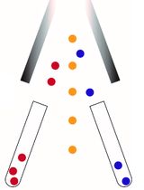
Figure 1. Experimental design for determination of transfection efficiency by flow cytometric method. The plasmid DNA was labeled with FITC by DNA label IT@ tracker. After transfection, cells were detected by flow cytometry. The FITC fluorochrome is used to detect intracellular levels of the transfected plasmid that has been labeled with FITC (Label IT tracker, green). The second fluorochrome is used to quantify expression of the target protein (by directly measuring fluorescence of the expressed protein if the target protein is fluorescent or by using a fluorescent-labeled antibody against the target protein, red). Either Q1+Q2 (DNA signal) or Q2+Q3 (protein signal) should be used as readouts of transfection efficiency.
Materials and Reagents
- 4 mm cuvettes (Gene Pulser cuvettes, Biorad, Hercules, CA)
- Cell lines of interest
- 293T cell line (ATCC, catalog number: A-498)
- Jurkat E6-1 (obtained through the NIH AIDS Reagent Program, Division of AIDS, NIH)
- Jurkat Clone E6-1 (from Dr. Arthur Weiss)
- pNL4-3, received from AIDS reagent program (#114, NIH)
- pUltraHot encoding mCherry (8,314 bp) was a kind gift of Jeff F. Miller, California Nano Systems Institute
- One ShotTM Stbl3TM Chemically Competent E. coli (Thermo Scientific, catalog number: C737303)
- Stellar bacteria (Clontech, catalog number: 636763)
- Dulbecco's modified Eagle medium (DMEM) (Thermo Scientific, catalog number: 11965-092)
- Roswell Park Memorial Institute (RPMI) 1640 medium (Thermo Scientific, catalog number: 61870127)
- Fetal bovine serum (FBS) (Omega Scientific, catalog number: FB-02)
- OptiMEM (Thermo Scientific, catalog number: 31985-070)
- Penicillin and streptomycin (Thermo Scientific, catalog number: 15-140-122)
- L-glutamine (Invitrogen, catalog number: 125030-081)
- Phosphate-buffered saline (PBS) (Omega Scientific, catalog number: FB-02)
- 4% paraformaldehyde (PFA) (Thermo Scientific, catalog number: J19943-K2)
- Tween 20 (Sigma, catalog number: P9416)
- PureLink HiPure Midiprep Kit (Thermo Scientific, catalog number: K210004)
- DNA Label IT® TrackerTM [Fluorescein isothiocyanate (FITC)] (Mirus, catalog number: MIR7025)
- TransIT-X2 (Mirus, catalog number: MIR6004)
- Jet Prime (Polyplus, catalog number: 114-07)
- Lipofectamine 2000 (Thermo Scientific, catalog number: 11668019)
- Fugene HD (Promega, catalog number: E2311)
- Quantum Molecules of Equivalent Soluble Fluorochrome (MESF) (Bangs Laboratories, catalog number: 647-B)
- Ghost Violet 450 Live/dead dye (Tonbo Biosciences, catalog number: 13-0863-T100)
- Anti-human immunodeficiency virus (HIV)-1 p24 monoclonal antibody (71-31) (NIH AIDS reagent program #530)
- Human IgG1 (Biolegend, catalog number: 409302)
- Mix-N-Stain CF647 dye (Biotium, catalog number: 92238)
- 10% FBS complete culturing medium for 293T (see Recipes)
- 10% FBS complete culturing medium for Jurkat cells (see Recipes)
- 0.02% Tween/PBS (see Recipes)
Equipment
- 37 °C, 5% CO2 humidified incubator (Thermoforma, model: 3110)
- Centrifuge (Thermo scientific, model: ST8)
- BioRad Gene Pulser Xcell electroporation system
- LSRII Fortessa flow cytometer (BD Biosciences, San Jose, CA, USA)
- Spectrophotometer (Thermo Fisher, NanoDropTM 2000)
Software
- Acquisition Software: BDFACSDiVaTM Software (BD Biosciences)
- Analysis Software: FlowJo version 10.6
Procedure
文章信息
版权信息
© 2019 The Authors; exclusive licensee Bio-protocol LLC.
如何引用
Mu, W., Homann, S., Hofmann, C., Gorin, A., Huynh, D., Yang, O. O. and Kelesidis, T. (2019). A Flow Cytometric Method to Determine Transfection Efficiency. Bio-protocol 9(10): e3244. DOI: 10.21769/BioProtoc.3244.
分类
细胞生物学 > 基于细胞的分析方法 > 病毒性感染
细胞生物学 > 单细胞分析 > 流式细胞术
您对这篇实验方法有问题吗?
在此处发布您的问题,我们将邀请本文作者来回答。同时,我们会将您的问题发布到Bio-protocol Exchange,以便寻求社区成员的帮助。