

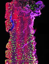
Immunofluorescence Staining of WT-1/Podocalyxin on Mouse Kidney Sections
免疫荧光标记小鼠肾脏切片中的WT-1/足糖萼

发布: 2019年04月20日第9卷第8期 DOI: 10.21769/BioProtoc.3210 浏览次数: 7053
评审: Anonymous reviewer(s)
参见作者原研究论文
The authors used this protocol in:

Nov 2017
Advertisement


Abstract
In glomerular diseases, podocytes are one type of cells involved in dysfunction of glomerular filtration. In these conditions, podocyte proteins expression change. Therefore, immunofluorescent staining of podocytes can be performed to visualize podocyte localization of proteins of interest. In this protocol, we detail a method to stain podocytes with a specific marker WT-1 (Wilms Tumor-1) for “in situ” podocyte analysis by immunofluorescent microscopy, more informative technique than other techniques (as Western blot).
Keywords: Immunofluorescence (免疫荧光)Background
In vivo, mature podocytes are glomerular epithelial cells that are normally terminally differentiated (Pavenstadt et al., 2003). Under pathological conditions, podocytes may undergo mitosis and proliferate. For example, crescentic glomerulonephritis is an exceptional condition where podocytes undergo a dysregulation of their differentiated phenotype and start to proliferate and migrate, resulting in the so-called extracapillary glomerulopathy. Several studies have shown involvement of podocytes in crescentic glomerulonephritis (Moeller et al., 2004).
Wilm’s tumor 1 (WT-1) is a zinc finger transcription factor present in nucleus of podocyte. Podocalyxin is a sialoglycoprotein constituting the glycocalyx of podocytes and endothelial cells in glomerulus. The expression of WT1 and podocalyxin are not decreased in early-stage of glomerular damage compared to proteins of slit diaphragm as podocin or nephrin. Moreover, WT1 is useful to evaluating podocyte number and density under different circumstances. The advantages to use podocalyxin staining are i) to analyze the glomerular morphology, ii) to quantify flocculus area to perform WT1+ cells / flocculus area ratio; iii) to ensure that WT1+ cells are podocytes and no other cells as parietal epithelial cells which could express WT1 in pathophysiological context.
Our protocol can be used in crescentic glomerulonephritis (Henique et al., 2017) but also in studies on different glomerulopathies as hypertensive nephropathy.
Materials and Reagents
- Slides Menzel-Gläser Superfrost® PLUS 25 x 75 x 1.0 mm (Thermo Fisher Scientific, catalog number: J1800AMNZ)
- Cover slip (Knittel Glass, 24 x 50 mm, catalog number: 023/40)
- PAP pen (DakoPen, Dako, catalog number: S2002) or equivalent
- Tissue Tek container/Staining Dish green (solvent-resistant) (Sakura, catalog number: SKU4456)
- Paper towels
- Aluminum foil
- 0.2 µm syringe filter (VWR, catalog number: 514-0060)
- Primary antibody
- Rabbit anti-Wilms Tumor Protein (WT-1) (dilution ratio 1:100, put 1 μl of antibody with 99 μl of blocking solution) (clone [CAN-R9(IHC)-56-2]) (Abcam, catalog number: ab89901)
- Biotinylated goat anti-mouse podocalyxin (dilution ratio 1:500, put 1 μl of antibody with 499 μl of blocking solution) (R&D, catalog number: BAF1556)
- Secondary antibody
- Alexa Fluor 594 donkey anti-rabbit (dilution ratio 1:400, put 1 μl of antibody with 399 μl of blocking solution) (Thermo Fisher Scientific, catalog number: A21207)
- Alexa Fluor 488 donkey-anti-goat (dilution ratio 1:400, put 1 μl of antibody with 399 µl of blocking solution) (Thermo Fisher Scientific, catalog number: A11055)
- Streptavidin, Alexa Fluor 488 (Thermo Fisher Scientific, catalog number: S32354)
- Xylene (Carlo Erba, catalog number: 528251)
- Ethanol absolute (Carlo Erba, catalog number: 3086072)
- Target retrieval solution (Dako, catalog number: S1699)
- Fluorescence Mounting Medium (Dako, catalog number: S3023)
- DAPI solution (1 mg/ml) (Thermo Fisher Scientific, catalog number: 62248)
- Clear Nail Polish (available at local drug store)
- Phosphate Buffer Saline 10x (Euromedex, catalog number: ET330)
- TritonTM X-100 (Sigma-Aldrich, catalog number: X100)
- Tween® 20 (Sigma-Aldrich, catalog number: P2287)
- Bovine serum albumin (BSA) (Sigma-Aldrich, catalog number: A2058)
- Sudan Black B (Acros organics, catalog number: 190160250)
- Tris (Euromedex, catalog number: 200923-A)
- NaCl (Euromedex, catalog number: 1112-A)
- Tris-buffered saline (TBS) (see Recipes)
- TBS-Tween (1x) (see Recipes)
- Permeabilization solution (see Recipes)
- Blocking solution (see Recipes)
- Sundan Black B solution (see Recipes)
Equipment
- Axioimager Z1 apoptome (Zeiss)
- Pressure cooker, any pressure cooker can be used, here Decloaking chamber (Biocare medical, model: DC2008)
- Benchtop orbital shaker (Stuart scientific gyro rocker, model: STR9)
- 4 °C refrigerator
Software
- Axiovision microscopy software (Version AxioVs40 V 4.8.2.0, Zeiss)
Procedure
文章信息
版权信息
© 2019 The Authors; exclusive licensee Bio-protocol LLC.
如何引用
Henique, C., Lenoir, O. and Tharaux, P. (2019). Immunofluorescence Staining of WT-1/Podocalyxin on Mouse Kidney Sections. Bio-protocol 9(8): e3210. DOI: 10.21769/BioProtoc.3210.
分类
细胞生物学 > 组织分析 > 组织成像
您对这篇实验方法有问题吗?
在此处发布您的问题,我们将邀请本文作者来回答。同时,我们会将您的问题发布到Bio-protocol Exchange,以便寻求社区成员的帮助。