- EN - English
- CN - 中文
On-demand Labeling of SNAP-tagged Viral Protein for Pulse-Chase Imaging, Quench-Pulse-Chase Imaging, and Nanoscopy-based Inspection of Cell Lysates
用于脉冲追踪成像,猝发脉冲追踪成像和基于纳米显微技术的细胞裂解物检测的融合SNAP病毒蛋白的按需标记
发布: 2019年02月20日第9卷第4期 DOI: 10.21769/BioProtoc.3177 浏览次数: 6977
评审: Vamseedhar RayaproluJolene RamseyKristin L. Shingler
Abstract
Advanced labeling technologies allow researchers to study protein turnover inside intact cells and to track the labeled protein in downstream applications. In the context of a viral infection, the combination of imaging and fluorescent labeling of viral proteins sheds light on their biological activity and interaction with the host cell. Initial approaches have fused fluorescent proteins such as green fluorescent protein (GFP) to the viral protein-of-interest. In contrast, self-labeling enzyme tags such as the commercial SNAP-tag, a modified version of human O6-alkylguanine-DNA-alkyltransferase, covalently link synthetic ligands, which users can add on demand. The first two protocols presented here build on previously published protocols for fluorescent labeling in pulse-chase and quench-pulse-chase experiments; the combination of fluorescent labeling with advanced light microscopy visualizes the dynamic turnover of the SNAP-tagged viral protein in intact mammalian cells. A third protocol also outlines how to inspect cellular lysates microscopically for detergent-resistant assemblies of the labeled viral protein. These protocols showcase the flexibility of the SNAP-based labeling system for tracking a viral protein-of-interest in live cells, intact fixed cells, and cell lysates. Moreover, the protocols employ recently developed commercial microscopes (e.g., Airyscan microscopy) that balance resolution, speed, phototoxicity, photobleaching, and ease-of-use.
Keywords: Fusion proteins (融合蛋白)Background
To better understand the role of a viral protein during the infectious life cycle, we have adapted existing strategies that facilitate the determination of intracellular protein location, the assessment of protein dynamics in intact cells, and the continued tracking of the viral protein after lysis of the host cell. The first two protocols build on previously published protocols for on-demand SNAP-labeling and the analysis of protein turnover (Bodor et al., 2012). In previous work, our laboratory has fused a viral protein-of-interest, nonstructural protein 3 (nsP3) of Chikungunya virus (CHIKV), to the SNAP-tag, a modified form of a 20-kDa monomeric DNA repair enzyme (Remenyi et al., 2017 and 2018). During SNAP-labeling, the addition of a synthetic O6-benzylguanine (BG) derivative results in a covalent bond between a reactive cysteine residue in the SNAP-tag and the BG-probe (Keppler et al., 2004a and 2004b).
We have also combined SNAP-labeling with a protocol for inspecting cell lysates via light microscopy, which enables visualization of detergent-resistant protein assemblies. Similar approaches have allowed researchers to detect stable granular assemblies of GFP-tagged stress-granule proteins in cell lysates (Jain et al., 2016; Wheeler et al., 2017). Stress granules are assemblies of RNA and protein (RNPs), which form under conditions of cellular stress (Kedersha et al., 2005). It is now possible to isolate the more stable stress granule core from both yeast and mammalian cells (Jain et al., 2016). SNAP-labeling offers an alternative way of tracking tagged viral proteins that may be present in similar subcellular assemblies. Hence, Protocol 3 may not only be useful to study the biochemical nature of viral proteins but also to track any cellular protein that resides in non-membranous organelles such as RNPs and stress granules. For example, integration of the SNAP-tag into the development of cell lines that produce fluorescently tagged stress granules (Kedersha et al., 2008) could increase experimental flexibility during dynamic and quantitative imaging of these cellular sub-compartments.
Our three protocols also take advantage of recently developed commercial imaging systems for multi-color fluorescence microscopy. We analyze labeled samples in Protocol 1 with live-cell imaging (Figure 1) and thus recommend following general procedures for controlling temperature, reducing phototoxicity, limiting photobleaching, and maintaining cell viability (Frigault et al., 2009). The chosen 2018 Nikon Ti-E2 system allowed us to image a large field-of-view, lessen focus drift with a proprietary Perfect Focus System (PFS), and record multiple positions with the motorized stage. Moreover, illumination with an LED and light exposures not exceeding 1 s allowed for gentler imaging compared to the typical imaging setup of a laser scanning confocal microscope.
For the analysis of labeled protein in Protocols 2 and 3, we chose a confocal imaging setup with proprietary ZEISS Airyscan technology (Figure 1), which is a commercial version of the ‘image scanning microscopy’ approach (Muller and Enderlein, 2010; Sheppard et al., 2013). Airyscan microscopy represents one of the recent innovations in fluorescence super-resolution microscopy, also referred to as nanoscopy (Li et al., 2018). The Airyscan technology improves system resolution with an improved detector design, which features a 32-channel detector array (Huff, 2015). With a new 2D super-resolution mode for Airyscan, this detection approach can now enhance resolution 2-fold while lowering the required fluorescence intensity to obtain high-quality images (Huff et al., 2017). The Airyscan system allowed us to stain our samples with fluorescent dyes from the SNAP product range (e.g., SNAP-Cell® 647-SiR and SNAP-Cell® TMR-Star) and detect them even at low levels during the recovery phase of the quench-pulse-chase protocol. Increased sensitivity also helped in the nanoscopy-based inspection of cellular lysate in Protocol 3 and allowed the visualization of granular structures made up of the SNAP-tagged viral protein. The combination of SNAP-based labeling and innovative detection via Airyscan has the potential to further bioimaging with higher resolution, sensitivity, and user-friendliness.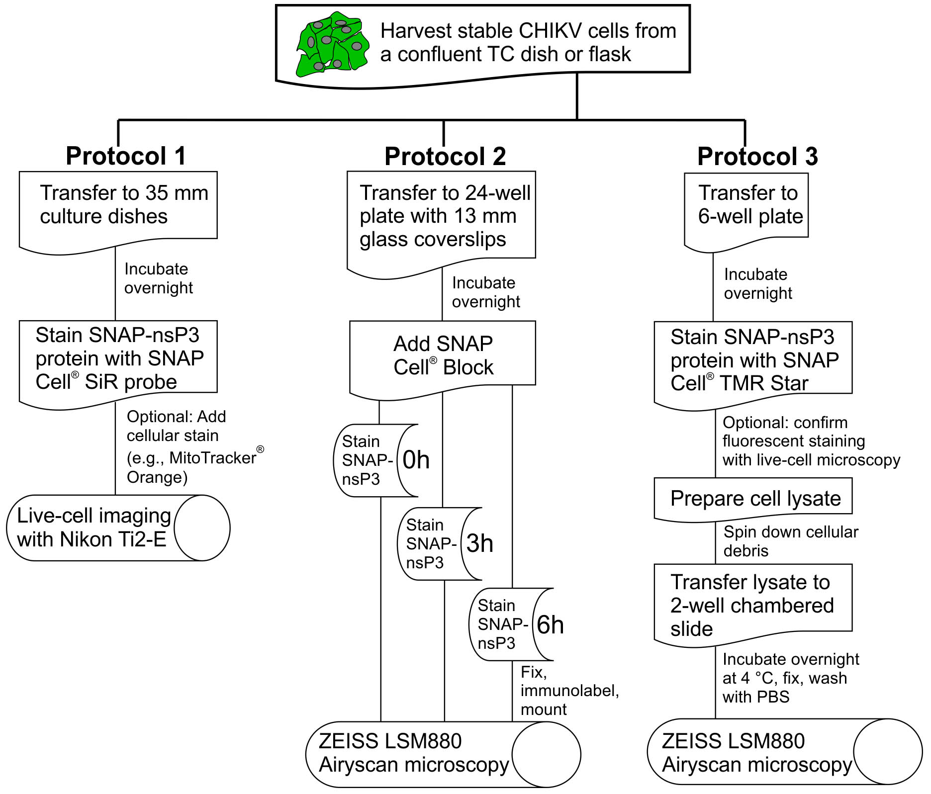
Figure 1. Overview of experimental protocols. This flowchart outlines the essential steps in Protocols 1, 2, and 3. For the definition of ‘stable CHIKV cells’, see Protocol 1, Procedure section.
Protocol 1: Pulse-chase experiments for long-term imaging with Nikon Ti2-E
Materials and Reagents
- 10-cm Petri dish (Corning, catalog number: 430167)
- Micropipette tips, serological pipettes, pipette aids, and microtubes for liquid handling
- TipOne® 1,000 µl XL (catalog number: S1122-1830)
- TipOne® 200 µl (catalog number: S1120-8810)
- TipOne® 20 µl filter tips (catalog number: S1120-1810) or SARSTEDT pipette tip 10 µl (catalog number: 70.1130.600)
- FisherbrandTM 5 ml serological pipets (catalog number: 13-676-10H)
- FisherbrandTM 10 ml serological pipets (catalog number: 13-676-10J)
- FisherbrandTM 25 ml serological pipets (catalog number: 13-676-10K)
- Drummond Pipet-Aid XL (catalog number: 4-000-205)
- Microtube 1.5 ml (SARSTEDT, catalog number: 72.690.001)
- Nunc 35-mm glass bottom (#1.5 or 0.16-0.19 mm thickness) dishes with 27 mm viewing area (Thermo Fisher Scientific, catalog number: 150682)
Notes:- Only open packages inside a biosafety cabinet and reseal any remaining dishes to avoid contamination.
- Use any dish or slide format that is compatible with an inverted microscope. Match the thickness of the glass bottom with the suggested thickness found on the microscope’s objective (typically #1.5 or 0.16-0.19 mm thickness). We prefer the dishes listed above because of their large viewing area. The cell lines used in this protocol can grow on glass substrates, but surface treatment (e.g., Poly-Lysine coating) may be necessary for other cell lines.
- HuH-7 cell line (Japanese Collection of Research Bioresources [JCBR], catalog number: JCBR 0403)
Note: HuH-7 is a well-differentiated hepatocyte-derived cellular carcinoma cell line. It was originally taken from a liver tumor in a Japanese male in 1982 (Nakabayashi et al., 1982). The cells used in the creation of this protocol were obtained from John McLauchlan (Centre for Virus Research, Glasgow). - A plasmid that encodes SNAPf®, a SNAP-tag protein (NEB, catalog number: N9183S; or as part of the SNAP-Cell® Starter Kit, NEB, catalog number: E9100S)
- For initial validation: plasmid that encodes pSNAPf-Cox8A control plasmid (also part of the SNAP-Cell® Starter Kit, NEB, catalog number: E9100S)
- Trypsin EDTA solution (Sigma, catalog number: T3924-500ML)
- Specific reagents for our cell line (for basic tissue culture), which we derived from the hepatoma cell line HuH-7
- Dulbecco's modified Eagle's medium (Sigma, catalog number: D6429-500ML)
- 100% Fetal Calf Serum (Gibco, catalog number: 10500-064)
- Gibco MEM Nonessential amino acids solution (100x), store at 4 °C up to 24 months from the date of manufacture (Thermo Fisher Scientific, catalog number: 11140050)
- Gibco HEPES (N-2-hydroxyethylpiperazine-N-2-ethane sulfonic acid) buffer (100x), store at 4 °C up to 24 months from the date of manufacture (Thermo Fisher Scientific, catalog number: 15630130)
- FluoroBrite DMEM, store at 4 °C up to 24 months from the date of manufacture (Thermo Fisher Scientific, catalog number: A1896701)
- Cell-permeable SNAP-Cell® 647-SiR (New England Biolabs, catalog number: S9102S)
Notes:- Package contains 30 nmol of the substrate. Resuspend with 50 µl of sterile dimethyl sulfoxide (DMSO, Fisher BioreagentsTM, catalog number: BP231-100) to make up a stock solution, which can be stored at -20 °C. We have used stock solutions that have been stored for up to 3 years. However, the manufacturer’s recommended shelf-life is three months dissolved in DMSO and two years dry.
- NEB also offers cell-impermeable SNAP probes (‘Cell Surface’ Probes); use these probes for studies of viral proteins that accumulate at the surface of host cells.
- Optional: Red fluorescent or orange fluorescent cellular dye (e.g., MitoTrackerTM Orange CMTMRos, Thermo Fisher Scientific, catalog number: M7510)
Note: To prepare a stock solution, dissolve lyophilized MitoTrackerTM probe in DMSO to a final concentration of 1 mM and store frozen and protected from light. - Molecular Probes Invitrogen ProLong Live Antifade Reagent, for live cell imaging (Thermo Fisher Scientific, catalog number: P36975)
Notes:- Store at 2-8 °C for short-term storage and ≤ -20 °C for long-term storage.
- According to the manufacturer’s instructions, use the product within 30 days when stored at 2-8 °C. When stored at ≤ -20 °C, the product is stable for at least six months with up to four freeze-thaw cycles.
- Glutamax-I (Thermo Fisher Scientific, catalog number: 35050061)
- Live cell imaging solution (see Recipes)
- Tissue culture medium containing serum (see Recipes)
Equipment
- Air-displacement micropipettes
- Starlab ErgoOne® 100-1,000 µl Single-Channel Pipette (catalog number: S7110-1000) or Gilson F123602 PIPETMAN Classic Pipet P1000 (Fisher Scientific, catalog number: 10387322)
- Starlab ErgoOne® 20-200 µl Single-Channel Pipette (catalog number: S7100-2200) or Gilson F123615 PIPETMAN Classic Pipet P100 (Fisher Scientific, catalog number: 10442412)
- Starlab ErgoOne® 2-20 µl Single-Channel Pipette (catalog number: S7100-0220) or Starlab ErgoOne® 0.1-2.5 µl Single-Channel Pipette (catalog number: S7100-0125) or Gilson F144801 PIPETMAN Classic Pipet P2 (Fisher Scientific, catalog number: 10635313) or Gilson F123600 PIPETMAN Classic Pipet P20 (Fisher Scientific, catalog number: 10082012)
- Equipment for basic cell culture techniques and aseptic procedures, i.e.,
- Biosafety cabinet (e.g., Thermo Scientific Holten Safe 2010 Model 1.2, catalog number: 8207071100)
- Humidified incubator set to 37 °C (Panasonic Incusafe, catalog number: MCO-20AIC)
- -20 °C freezer (Labcold, catalog number: RLCF1520)
- Water bath (e.g., Clifton, catalog number: NE2-8D; however, any water bath set to 37 °C may be used)
- Table-top microcentrifuge, 4 °C to room temperature (RT), max speed ≥ 12,000 x g (Eppendorf, catalog number: 5424R)
Note: For spinning down precipitate that may form during extended storage of SNAP probes. - Live-cell imaging microscope for long-term imaging. For example, a Nikon Ti2-E inverted widefield microscope (for a similar setup, contact your local Nikon representative to create a customized order for the microscope system)
- Nikon Ti2-E inverted microscope stand
- Motorized stage (standard Ti2 encoded motorized XY stage)
- Lumencor Spectra X LED light source
- CFI Plan Apo Lambda 60x oil/1.4 NA objective
- Photometric Prime 95B sCMOS monochrome camera
- Semrock 32 mm filter (Green) for Nikon Ti2-E: GFP-4050B Filter cube with Ex 466/40 single-band bandpass filter (product code: FF01-466/40-25), DM495 dichroic beamsplitter (product code: FF495-Di03-25x36), BA525/50 single-band bandpass filter (product code: FF03-525/50-25)
- Semrock 32 mm filter (Red) for Nikon Ti2-E: Cy3-4040C Filter cube with Ex 531/40 single-band bandpass filter (product code: FF01-531/40-25), Dichroic Mirror DM 562 (product code: FF562-Di03-25x36, Barrier Filter: BA 593/40 (product code: FF01-593/40-25)
- Semrock 32 mm filter (Far-Red) for Nikon Ti2-E: Cy5-4040C Filter cube with Ex 628/40 single-band bandpass filter (product ID: FF02-628/40-25), Single Band Emitter (product ID: FF01-692/40-25), Single Band Dichroic (product ID: FF660-Di02-25x36)
- Okolab stage top incubator, catalog number: H301-NIKON-NZ100/200/500-N. Set to 37 °C with 5% CO2 (any manufacturer and model that can be mounted on a Nikon Ti2 motorized stage will do)
- Perfect Focus System (PFS)
- The above list is not exhaustive but rather lists the essential components that have been useful in the creation of Protocol 1. Consult with your local Nikon representative for the remaining components of a customized microscope system.
- We prefer to image cells in open tissue-culture dishes with a coverslip-like glass bottom, which necessitates an inverted microscope stand.
- As Protocol 1 is a powerful approach for long-term imaging of the labeled protein, it is important to keep cells in focus and correct for focus drift caused by thermal and mechanical conditions. This live-cell imaging system included Nikon’s fourth-generation Perfect Focus System (PFS), which monitors the partial reflection of a low-power infrared laser beam on the interface between the dish’s glass bottom and liquid media above. This feature provides continuous and real-time focus drift correction.
- A large (25 mm x 25 mm) field of view (FOV) enables increased data throughput and capture of additional cells that would be outside the normal FOV.
- We preferred an LED light source over laser-based illumination. LED illumination limited photobleaching and phototoxicity; ‘gentle’ imaging was essential during acquisition of five-dimensional datasets (5-D: multi-color 3-D Z-stacks over time).
- A motorized microscope stage and the ability to record multiple positions during one experiment further increased the throughput of the imaging system.
Software
- Nikon NIS Elements AR imaging software
Nikon NIS Elements AR imaging software controls all components of the microscope. Use NIS Elements to set-up parameters for acquisition of 5-D datasets. Users can also create rendered videos within NIS Elements and save individual frames of 5-D data. For our widefield images, we also used a Richardson-Lucy algorithm (set to 10 iterations) to deconvolve datasets within the Elements software.
Procedure
文章信息
版权信息
© 2019 The Authors; exclusive licensee Bio-protocol LLC.
如何引用
Remenyi, R., Li, R. and Harris, M. (2019). On-demand Labeling of SNAP-tagged Viral Protein for Pulse-Chase Imaging, Quench-Pulse-Chase Imaging, and Nanoscopy-based Inspection of Cell Lysates. Bio-protocol 9(4): e3177. DOI: 10.21769/BioProtoc.3177.
分类
细胞生物学 > 细胞成像 > 荧光
细胞生物学 > 细胞成像 > 活细胞成像
您对这篇实验方法有问题吗?
在此处发布您的问题,我们将邀请本文作者来回答。同时,我们会将您的问题发布到Bio-protocol Exchange,以便寻求社区成员的帮助。
Share
Bluesky
X
Copy link


