

Heterochronic Phenotype Analysis of Hypodermal Seam Cells in Caenorhabditis elegans
秀丽隐杆线虫下皮干细胞的异时表型分析
发布: 2019年01月05日第9卷第1期 DOI: 10.21769/BioProtoc.3132 浏览次数: 6562
评审: Manish ChamoliSanjib GuhaAnonymous reviewer(s)
参见作者原研究论文
The authors used this protocol in:
Mar 2018
Advertisement


Abstract
Heterochrony refers to changes in the timing of developmental events, and it is precisely regulated in the organisms by the heterochronic genes such as C. elegans lin-4 and let-7. Mutations in these genes cause precocious or retarded development of certain cell lineages. With well-defined cell lineages, C. elegans is one of the best model systems to study heterochronic genes, since the subtle changes in the development of cell lineages can be easily identified. Among the different cell types in C. elegans, hypodermal seam cells and their lineages are well known to be maintained by lin-14, whose expression level is regulated by two miRNA genes, lin-4 and let-7, at the larval stages. Therefore, analyzing the heterochronic phenotype of hypodermal seam cells in C. elegans could yield detailed insights into the status of the miRNA pathway. Here we describe the assay protocol to analyze the heterochronic phenotypes of C. elegans hypodermal seam cells, which can be used as a reliable method to study the miRNA pathway.
Keywords: C. elegans (秀丽隐杆线虫)Background
Caenorhabditis elegans is a transparent nematode which is found in the soil. It was introduced as a new model organism by Sydney Brenner in the 1960s to study neural development. Since then, it has been extensively studied because of the simple anatomy, the easy cultivation, and the rapid growth. The reproductive life cycle of C. elegans, which takes only three days, consists of the embryonic stage, four larval stages (L1-L4), and the adult stage. After 14 h of embryogenesis, C. elegans grows in size during the four larval stages which are divided by each molt, then it reaches the adult stage which can produce the next generation. In unfavorable conditions, C. elegans larvae can choose the alternative developmental pathway, so-called “dauer”, which can survive a few months in the adverse conditions. When the environment becomes favorable, C. elegans exits the dauer stage and develops into the L4 stage.
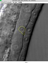
In C. elegans, most hypodermal seam cells have the characteristic of stem cells. In each molt, they divide into two daughter cells; the anterior daughter cell fuses with hyp7, which is the major hypodermis, and the posterior daughter cell continues to divide like the stem cells until they terminally differentiate at the L4 stages. Thus, ten hypodermal seam cells at L1 stage generate sixteen hypodermal seam cells at the end of the L4 stage in each side (Figure 1) (Altun and Hall, 2009).
Functionally, hypodermal seam cells are important for the formation of stage-specific cuticles that are composed of various collagen proteins (Thein et al., 2003). They also produce cuticular alae, which are the protruding ridges extending longitudinally along the two sides of the animal over the seam cells (Singh and Sulston, 1978). Alae are produced only in the L1 stage, dauer larva, and adult. Therefore, adult alae are commonly used as an indicator which shows terminal differentiation of hypodermal seam cells. For example, lin-4 or let-7 loss-of-function mutant, which shows the retarded development of hypodermal seam cells, has the alae defects in adult stages, while lin-14 loss-of-function mutant, which shows the precocious development of hypodermal seam cells, has alae in the L3 stage (Hong et al., 2000).
The cell fate of a hypodermal seam cell is regulated by lin-14, whose expression is high in L1 animals, and decreases by the L2 stage (Ruvkun and Giusto, 1989). The lin-14 expression is regulated by well-characterized miRNAs lin-4 and let-7 (Ambros, 1989; Reinhart et al., 2000). It is known that lin-4 and let-7 bind the 3’ untranslated region of lin-14 and downregulate its expression at the larval stages (Lee et al., 1993, Slack et al., 2000). When the expression level of lin-14 is not decreased in the larval stages, hypodermal seam cells abnormally proliferate, generating retarded phenotypes in lin-14 gain-of-function mutants and lin-4 loss-of-function mutants. 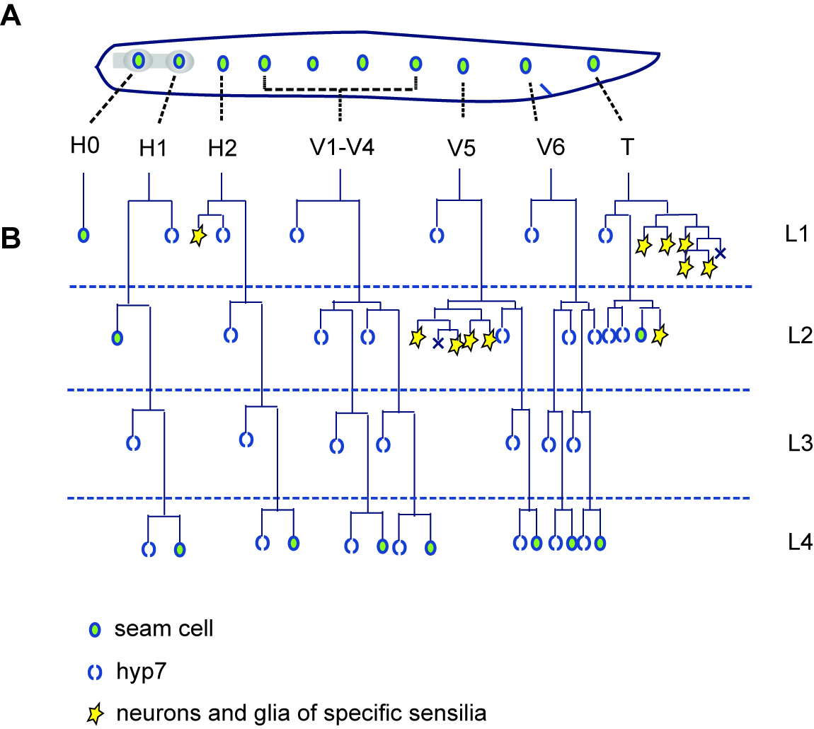
Figure 1. Hypodermal seam cell lineage in C. elegans. A. Ten hypodermal seam cells at the L1 stage. These cells generate major hypodermis hyp7, hypodermal seam cells, neurons and glia throughout the development. B. Cell division patterns of hypodermal seam cells in each molt. Most of the hypodermal seam cells generate one terminally differentiated daughter cell and one stem-cell-like seam cell in each division. By the L4 stage, sixteen of hypodermal seam cells are generated in each lateral side.
LIN-14 is a transcription factor that regulates its target gene expressions. One of the target genes of LIN-14 transcription factor is a cell cycle inhibitor cki-1. Inactivation of cki-1. results in the division of the vulva precursor cell (VPC) during the L2 stage. Therefore, decreasing lin-14 expression via lin-4 miRNA is important for the development of vulva as well as the hypodermal seam cell.
miRNAs are small, non-coding RNAs that are produced by a series of RNA-processing steps from their precursors known as pri-miRNAs. The pri-miRNAs are generated by transcription via RNA polymerase II (Bartel, 2004; Lee et al., 2004) and then cleaved by the RNase III endonuclease Drosha to produce pre-miRNAs in the nucleus (Bracht et al., 2004; Lee et al., 2002). These pre-miRNAs are then transported into the cytoplasm by Exportin-5 (Yi et al., 2003). In the cytoplasm, the pre-miRNA is further processed by Dicer to produce the mature 20-25 nt miRNA (Bernstein et al., 2001; Grishok et al., 2001). The mature miRNA functions as a guide to recruit RNA-induced silencing complex (RISC), which is composed of the miRNA:mRNA duplex, Argonaute, and other proteins (Hammond et al., 2001; Carmell et al., 2002; Caudy et al., 2002; Mourelatos et al., 2002; Caudy et al., 2003). In the RISC complex, Argonaute cleaves the target mRNA when it is activated. In C. elegans, there are about 24 Argonaute proteins (Bartel, 2004; Carmel et al., 2002). Among them, ALG-1 and AGL-2 are appeared to be important for downregulating of lin-4 and let-7 targets, because alg-1 and alg-2 mutants share the phenotypes with lin-4 and let-7 mutants. (Grishok et al., 2001).
Here we describe the assay protocols to analyze the heterochronic phenotypes of hypodermal seam cells in C. elegans. These assays are useful for the analysis of the miRNA pathway by taking advantage of the fact that hypodermal seam cell fate is regulated by the well-characterized miRNAs, lin-4 and let-7 (Zhang et al., 2018).
Materials and Reagents
- 60 mm Petri dishes (Fisher Scientific, FisherbrandTM, catalog number: AS4051)
- 100 mm Petri dishes (Fisher Scientific, FisherbrandTM, catalog number: FB0875712)
- Syringe Filter Unit, 0.22 μm (Millipore Sigma, Millex®-GV, catalog number: SLGV033RS)
- Frosted microscope slides 25 x 75 x 1.0 mm (Fisher Scientific, FisherbrandTM, catalog number: 12-552-3)
- Microscopic cover glass 18 x 18 mm (Fisher Scientific, FisherbrandTM, catalog number: 12-542A)
- C. elegans wild type: N2 Bristol strain from C. elegans Genetic Center
- JR672 wIs54 [Pscm::gfp] V; wIs54 is the integration allele of the seam cell-specific transcriptional GFP reporter that is expressed in all of the seam cells at all developmental stages (Terns et al., 1997; Koh and Rothman, 2001)
- E. coli OP50-1: streptomycin resistant strain from C. elegans Genetic Center
- Levamisole (Sigma-Aldrich, catalog number: L9756)
- NaCl (Fisher Scientific, Fisherbrand, catalog number: S271, CAS 7647-14-5)
- Agar, Bacteriological, Ultrapure (Thermo Scientific, catalog number: J10906, CAS 9002-18-0)
- Peptone (BD Bioscience, BD BactoTM, catalog number: 211677)
- Calcium Chloride hexahydrate (Sigma-Aldrich, catalog number: 442909)
- Magnesium sulfate (Sigma-Aldrich, catalog number: 208094)
- Potassium phosphate dibasic (Sigma-Aldrich, catalog number: P3786)
- Potassium phosphate monobasic (Sigma-Aldrich, catalog number: P0662)
- Cholesterol (Sigma-Aldrich, catalog number: C75209)
- Pure alcohol 200 proof (Pharmco products, catalog number: 111000200)
- Tryptone (BD Bioscience, BD BactoTM, catalog number: 211705)
- Yeast Extract (BD Bioscience, BD BactoTM, catalog number: 212750)
- Nail polish
- Nematode Growth Media (NGM) Agar (see Recipes)
- 1 M CaCl2 (see Recipes)
- 0.5 M MgSO4 (see Recipes)
- 1 M Potassium phosphate (pH 6) (see Recipes)
- 5 mg/ml cholesterol (see Recipes)
- LB agar (see Recipes)
- LB (see Recipes)
- OP50-1 culture (see Recipes)
Equipment
- Amsco® Century SV-120 Scientific Prevacuum Sterilizer (STERIS)
- 4 L flask
- Stir bar
- Stir plate
- PourBoy® 4 Sterile Media Dispenser (TritechTM Research)
- Home-made cell spreader (made with glass Pasteur pipets by bent by heating, spreader size is less than 1 inch.)
- 37 °C incubator (VWR, model: 1535)
- Incubator Shaker (Eppendorf, New Brunswick Scientific, model: I2500, catalog number: M1284-0000)
- 20 °C incubator for C. elegans culture (Intellus control system) (PERCIVAL, model: I-36NL)
- A home-made worm picker with a 5¾ glass Pasteur pipet (Fisher Scientific, FisherbrandTM, catalog number: 13-678-6A) and a platinum wire (Scientific Instrument Services, Inc., catalog number: W414)
- Fluorescent Stereo Microscope (Leica Microsystems, model: Leica M165FC) with Leica PLANAPO 2.0x objective lens
- Confocal laser scanning microscopy platform (Leica Microsystems, model: Leica TCS SP8) with Leica CTR6500 electronics box
Software
- GraphPad Prism 7.0a (GraphPad Software, Inc., www.graphpad.com)
- Leica Application Suite Advanced Fluorescence (Leica Microsystems)
Procedure
文章信息
版权信息
© 2019 The Authors; exclusive licensee Bio-protocol LLC.
如何引用
Ji, Y. J. and Wang, J. (2019). Heterochronic Phenotype Analysis of Hypodermal Seam Cells in Caenorhabditis elegans. Bio-protocol 9(1): e3132. DOI: 10.21769/BioProtoc.3132.
分类
发育生物学 > 细胞生长和命运决定 > 分化
神经科学 > 细胞机理 > 神经元命运
细胞生物学 > 基于细胞的分析方法 > 异时表型
您对这篇实验方法有问题吗?
在此处发布您的问题,我们将邀请本文作者来回答。同时,我们会将您的问题发布到Bio-protocol Exchange,以便寻求社区成员的帮助。