

Eimeria vermiformis Infection Model of Murine Small Intestine
蠕形艾美耳球虫对小鼠小肠的感染模型
(*contributed equally to this work) 发布: 2018年12月20日第8卷第24期 DOI: 10.21769/BioProtoc.3122 浏览次数: 6661
评审: Ruth A. FranklinMigla MiskinyteBenoit Stijlemans
参见作者原研究论文
The authors used this protocol in:
Jun 2018
Advertisement


Abstract
Eimeria vermiformis is a tissue specific, intracellular protozoan that infects the murine small intestinal epithelia, which has been widely used as a coccidian model to study mucosal immunology. This mouse infection model is valuable to investigate the mechanisms of host protection against primary and secondary infection in the small intestine. Here, we describe the generation of an E. vermiformis stock solution, preparation of sporulated E. vermiformis to infect mice and determination of oocysts burden. This protocol should help to establish a highly reproducible natural infection challenge model to study immunity in the small intestine. The information obtained from using this mouse model can reveal fundamental mechanisms of interaction between the pathogen and the immune response, e.g., provided by intraepithelial lymphocytes (IEL) at the basolateral site of epithelial cells but also a variety of other immune cell populations present in the gut.
Keywords: Eimeria vermiformis (蠕形艾美耳球虫)Background
Infections of the small intestine are widespread and can cause significant morbidity and mortality worldwide (Munot and Kotler, 2016). Children and the elderly, due to their weaker immune system, suffer the worst consequences. Gaining a better understanding of how the small intestinal immune system functions is therefore of prime importance. However, many of the pathogens used in mouse models are not natural to the host, can cause systemic inflammation or can persist in the tissues for long periods. This can result in uncharacteristic induction of the immune response and inadequate development of protective immunity.
Eimeria vermiformis is a tissue specific, intracellular single cell protozoan, which infects the small intestinal epithelia. Unlike the related pathogen used in infection models, Toxoplasma gondii, this is a natural, self-limiting, murine parasite, which induces a strong immune response and establishes long-lasting protection against subsequent infections. Furthermore, E. vermiformis is monoxenous, its life cycle is completed without the need of a second host species (see Figure 1). It is therefore an ideal pathogen to study the mechanisms of host protection against primary and secondary infection at the small intestinal barrier.
Mice infected with E. vermiformis excrete a non-infectious, unsporulated form of oocysts into the environment. Once in contact with oxygen and moisture, in a process which takes between 2 to 7 days, these oocysts mature into an infectious, sporulated form, which, upon ingestion, disseminates the infection to other animals (Fayer, 1980).
The E. vermiformis infection model has been used in the past and has yielded valuable information in areas such as the role of αβ and γδ T cells that line the small intestinal epithelia (Roberts et al., 1996) and the influence of age of the host in the development of protective immune responses in the intestine (Ramsburg et al., 2003). More recently we have used the E. vermiformis model to highlight the role of mitochondria in controlling the activation state of epithelial-resident T lymphocytes (Konjar et al., 2018).
Here, we present an adapted version of the original protocol used by Long et al. (1976) to purify and sporulate E. vermiformis. We describe in detail the method used to propagate the parasite, infect mice and accurately assess parasite load.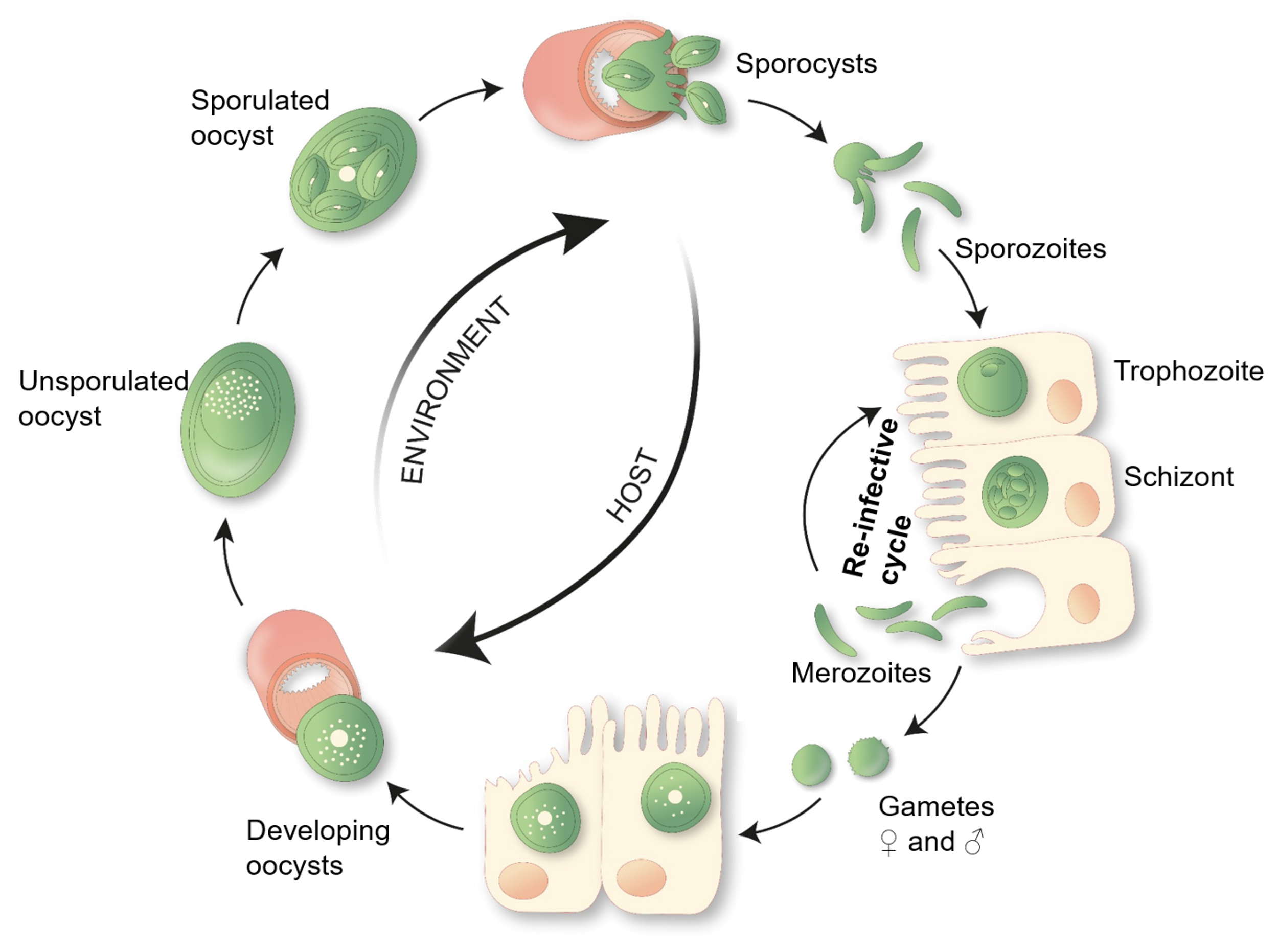
Figure 1. E. vermiformis life cycle. A single sporulated oocyst can initiate an infection when swallowed by its respective host, the mouse. Each mature oocyst holds four sporocysts, each sporocyst gives rise to two sporozoites, which can infect the enterocytes. Inside the host cell, the parasite develops via trophozoite and schizont stages into merozoites. Merozoites can re-infect the epithelial cells and start a new cycle. Eimeria typically undergoes three rounds of asexual multiplication whereupon merozoites give rise to gametes. Male and female gametes can fuse to form an oocyst, protected by a multi-layered cell wall making them highly resistant to environmental pressures. These oocysts shed with the feces. Unsporulated oocysts undergo sporulation upon contact with oxygen and moisture, undergoing meiosis, which takes between 2 to 7 days.
Materials and Reagents
- Millex®-GP Filter Unit: Millipore Express® PES membrane 0.22 μm (Merk Millipore Ltd., catalog number: SLGP033RS)
- Fine mesh metal sieve (no specific brand) (2x)
- Aluminum foil (no specific brand)
- 50 ml centrifuge tube, CentriStarTM cap, polypropylene, RNase-/DNase-free, Nonpyrogenic (Corning Science Mexico, catalog number: 430921)
- 15 ml centrifuge tube, CentriStarTM cap, polypropylene, RNase-/DNase-free, Nonpyrogenic (Corning Science Mexico, catalog number: 430791)
- 2 ml microcentrifuge tube graduated natural RNase-/DNase-free (Fisher Scientific, catalog number: 05-408-138)
- 1,000 μl Pipette tips (no specific brand)
- 200 μl Pipette tips (no specific brand)
- Fine sand, e.g., for playground and sand pits (Sand can be autoclaved for use in biological containment facilities)
- C57BL6/J mouse strain and/or strain and lines of interest
- Eimeria vermiformis (not commercially available)
- Sterile tap water
- Sodium hypochlorite 14% C12 in aqueous solution (VWR CHEMICALS, catalog number: 27900.365) (Room temperature)
- Sodium chloride (NaCl) (Merck KGaA, catalog number: 1. 06404.1000) (Room temperature)
- Potassium dichromate (Cr2K2O2) (VWR, catalog number: 26784.297)
- Fridge (no specific brand)
Equipment
- Fuchs-Rosenthal Counting Chamber (MARIENFELD, catalog number: 0640410)
- Two-chamber McMaster Counting Chamber (CHALEX, LLC: www.vetslides.com)
- Funnel (no specific brand)
- 1 L Beaker (no specific brand)
- P1000 Pipette (no specific brand)
- P200 Pipette (no specific brand)
- Magnetic stirrer (VELP Scientifica, model: F20500162)
- Fume hood (Burdinola, model: OR-ST-1500)
- Aquarium pump (no specific brand, e.g., 1 W silent pump)
- Centrifuge (Eppendorf AG, 22331 Hamburg, model: 5810 R)
- Grant-Bio Vortex, PV-1 (Grant Instruments Ltd)
- Microscope (Nikon, Japan, model: SE)
- Inverted microscope (EU importer-Carl Zeiss Microscopy GmbH, model: Primovert, catalog number: 415519-1100-000)
Procedure
文章信息
版权信息
© 2018 The Authors; exclusive licensee Bio-protocol LLC.
如何引用
Figueiredo-Campos, P., Ferreira, C., Blankenhaus, B. and Veldhoen, M. (2018). Eimeria vermiformis Infection Model of Murine Small Intestine. Bio-protocol 8(24): e3122. DOI: 10.21769/BioProtoc.3122.
分类
免疫学 > 粘膜免疫学 > 上皮
微生物学 > 体内实验模型 > 原生动物
细胞生物学 > 模式生物培养 > 保存
您对这篇实验方法有问题吗?
在此处发布您的问题,我们将邀请本文作者来回答。同时,我们会将您的问题发布到Bio-protocol Exchange,以便寻求社区成员的帮助。