

Relative Quantitation of Polymerized Actin in Suspension Cells by Flow Cytometry
利用流式细胞术相对定量分析悬浮细胞中的聚合肌动蛋白
发布: 2018年11月20日第8卷第22期 DOI: 10.21769/BioProtoc.3094 浏览次数: 6285
评审: Ralph Thomas Boettchertakashi nishinaPavan Vedula
参见作者原研究论文
The authors used this protocol in:

Jun 2017
Advertisement


Abstract
The amount of polymerized actin within a cell can vary widely due to natural processes and/or experimentally induced perturbations. We routinely use this protocol to measure relative polymerized actin content between cell populations by staining cells in suspension with fluorescent phalloidin, then measuring total cell fluorescence using flow cytometry. Although developed for human cells, we have easily adapted this method for use with diverse eukaryotic cell types.
Keywords: Actin (肌动蛋白)Background
Biologists have long studied actin polymers using biochemical assays and fluorescence microscopy. The latter has been facilitated by the discovery of phalloidin, a small molecule toxin produced by poisonous mushrooms that stabilizes actin structures by specifically binding adjacent monomers within actin polymers (Vandekerckhove et al., 1985). Fluorescent phalloidin conjugates stain complex actin structures and facilitate detailed visualization by fluorescence microscopy. However, quantitating this fluorescence can be painstaking, particularly when tracing population-level changes. In contrast, flow cytometry quantitates the total fluorescence intensity of thousands of cells per minute.
Here we describe how we estimate differences in the amount of actin polymer between cell populations by staining cells with fluorescent phalloidin, measuring the total cell fluorescence of individual cells using flow cytometry, and finally comparing the fluorescence values of the populations. It is important to note that this approach is comparative and cannot be used to measure the absolute concentrations of cellular actin polymer. This approach requires cells to be in suspension; if your cells require adhesion for proper actin polymerization, we suggest using quantitative fluorescence microscopy as described above.
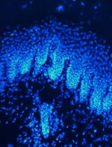
Although rapid and quantitative, flow cytometry provides no information about cell morphology. Therefore, we routinely save an aliquot of stained cells for visualization using fluorescence microscopy. We highly recommend doing this to ensure proper preservation of your structures of interest (Figure 1). Fluorescence microscopy can also reveal variation in experimental samples that are not apparent by flow cytometry.
This protocol is optimized for differentiated HL-60 cells that can be stimulated with fMLP to induce rapid actin polymerization (Figure 1, for additional information about these cells, see Fritz-Laylin et al., 2017), it can be modified for use with a wide variety of cell types by testing a variety of fixation conditions and buffers. For example, we have adapted this protocol for chytrid fungi (Batrachochytrium dendrobatidis), and the amoeba Naegleria gruberi (unpublished). To do this, we chose conditions that: 1) best preserve the overall morphology reasonably similar to live cells as visualized using DIC or phase contrast microscopy; 2) keep the polymerized actin intact through fixation; 3) when imaged by fluorescence microscopy, stain the appropriate structures. For information on buffers and fixatives (see Note 1) commonly used to preserve actin structures, we recommend the methods resource Fluorescence Procedures for the Actin and Tubulin Cytoskeleton in Fixed Cells from the Mitchison lab at Harvard University (L. Cramer and A. Desai).
Figure 1. An example of actin polymerization in response to cell stimulation. Maximum projection of AlexaFluor® 488 phalloidin stained HL-60 cells before and after treatment with 20 nM fMLP, which induces actin polymerization and results in brighter cells.
Materials and Reagents
- Standard uncoated 1.5 ml microcentrifuge tubes (USA Scientific, catalog number: 1615-5510)
- 15 and 50 ml conical tubes (any supplier)
- FACS tubes: sterile 5 ml polystyrene round-bottom tubes with a cell-strainer cap for filtering samples prior to flow cytometry (BD Falcon, catalog number: 352235), external filters are also available
Note: Check your cytometer requirements and/or your flow cytometry facility's recommendation for tubes. - Micropipette tips (Pipette.com, catalog numbers: UE-1000, UE-200, UE-10)
- Aluminum foil
- Razor blades
- Differentiated HL-60 cells (grown in medium RPMI supplemented with 10% Fetal Bovine Serum, and treated with 1.3% DMSO for 5 days; for additional details see Fritz-Laylin et al., 2017), or any other cell type for which you wish to quantitate relative amounts of polymerized actin
- RPMI (Gibco, catalog number: 11875)
- AlexaFluor® 488 conjugated Phalloidin (Thermo Fisher Scientific, catalog number: A12379) (see Note 3)
- Sterile Milli-Q water
- TWEEN-20 (Sigma-Aldrich, catalog number: P9416)
- Triton X-100 (Sigma-Aldrich, catalog number: X100)
- MES (Sigma-Aldrich, catalog number: M3671)
- KCl (Fisher Scientific, catalog number: P217-100)
- MgCl2 (Sigma-Aldrich, catalog number: M9272)
- EGTA (Sigma-Aldrich, catalog number: E4378)
- NaCl (Fisher Scientific, catalog number: BP3581)
- Na2HPO4 (Sigma-Aldrich, catalog number: 795410)
- KH2PO4 (Sigma-Aldrich, catalog number: P0662)
- BSA (Fisher Scientific, catalog number: BP1600-100)
- 16% Paraformaldehyde (Electron Microscopy Sciences, catalog number: 15710)
- DMSO (Sigma-Aldrich, catalog number: D2650)
- fMLP (Sigma-Aldrich, catalog number: 47729)
- Culture medium for HL-60 cells (see Recipes)
- Fixation Buffer (see Recipes)
- Cytoskeleton Buffer (see Recipes)
- Permeabilization Buffer (see Recipes)
- Staining solution (see Recipes)
- PBS (10x) (see Recipes)
- Wash solution (see Recipes)
Equipment
- Pipettes (Eppendorf, P1000, P100, P20, P1)
- Microcentrifuge with rotor suitable for 1.5 ml centrifuge tubes (e.g., Eppendorf, model: 5415)
- Flow cytometer (e.g., FACSCalibur analyzer [BD, model: FACSCaliburTM] or LSRFortessa Dual [BD, model: LSRFortessaTM])
- Microscope equipped for fluorescence and transmitted light (we prefer phase and/or DIC) (e.g., Nikon, model: Eclipse Ti-E)
- Incubator for growing cells, for HL-60 cells you will need a chamber with continuous 37 °C and 5% CO2
- Autoclave
- Microscope filters approprisate for the phalloidin conjugate of your choice (see Note 3)
- Standard hemocytometer (e.g., Sigma-Aldrich, catalog number: Z359629)
- Vortexer
- -20 °C Freezer
Software
- FACSDiva, BD (Becton, Dickinson & Company)
- FlowJo version 10, BD (Becton, Dickinson & Company)
- GraphPad Prism version 7, GraphPad Software
Procedure
文章信息
版权信息
© 2018 The Authors; exclusive licensee Bio-protocol LLC.
如何引用
Readers should cite both the Bio-protocol article and the original research article where this protocol was used:
- Kakley, M. R., Velle, K. B. and Fritz-Laylin, L. K. (2018). Relative Quantitation of Polymerized Actin in Suspension Cells by Flow Cytometry. Bio-protocol 8(22): e3094. DOI: 10.21769/BioProtoc.3094.
- Fritz-Laylin, L. K., Lord, S. J. and Mullins, R. D. (2017). WASP and SCAR are evolutionarily conserved in actin-filled pseudopod-based motility. J Cell Biol 216(6): 1673-1688.
分类
细胞生物学 > 细胞染色 > 蛋白质
生物化学 > 蛋白质 > 定量
您对这篇实验方法有问题吗?
在此处发布您的问题,我们将邀请本文作者来回答。同时,我们会将您的问题发布到Bio-protocol Exchange,以便寻求社区成员的帮助。