

Single-molecule Fluorescence in situ Hybridization (smFISH) for RNA Detection in Adherent Animal Cells
利用单分子荧光原位杂交进行贴壁动物细胞中RNA的检测

发布: 2018年11月05日第8卷第21期 DOI: 10.21769/BioProtoc.3070 浏览次数: 34618
评审: Samantha E. R. Dundontakashi nishina
参见作者原研究论文
The authors used this protocol in:

Nov 2017
Advertisement


Abstract
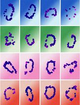
Transcription and RNA decay play critical roles in the process of gene expression and the ability to accurately measure cellular mRNA levels is essential for understanding this regulation. Here, we describe a single-molecule fluorescent in situ hybridization (smFISH) method (as performed in Haimovich et al., 2017) that detects single RNA molecules in individual cells. This technique employs multiple single-stranded, fluorescent labeled, short DNA probes that hybridize to target RNAs in fixed cells, allowing for both the quantification and localization of cytoplasmic and nuclear RNAs at the single-cell level and single-molecule resolution. Analyzing smFISH data provides absolute quantitative data of the number of cytoplasmic (“mature”) mRNAs, the number of nascent RNA molecules at distinct transcription sites, and the spatial localization of these RNAs in the cytoplasm and/or nucleoplasm.
Keywords: mRNA (mRNA)Background
Regulation of gene expression is one of the key determinants of cell fate and behavior. A major parameter of gene expression is mRNA level, which is determined by the rates of transcription and degradation. Therefore, measuring mRNA levels, as well as transcription and decay rates for particular transcripts (or all transcripts) has been the focus of numerous research projects.
Common molecular biology techniques, such as reverse transcription-PCR (RT-PCR), Northern blot analysis or RNA sequencing (RNA-Seq), typically require RNA extraction from the entire cell population. However, the results provide only a relative measure of mRNA content for the entire cell population, with a loss of single cell information. Single-cell RNA-Seq can provide more insight on the cell-to-cell variability of transcript levels. However, the current lower limit of detection is ~10 molecules/cell for a given RNA transcript (Svensson et al., 2017). RNA localization studies have shown that the spatial distribution of RNA in the cell can play a pivotal role in its function (Buxbaum et al., 2015), but the above-described methods lose that information in the process.
Single-molecule Fluorescence in situ Hybridization (smFISH) overcomes these limitations. In this method, the cells are first fixed and permeabilized. Then the cells are hybridized with a set of probes consisting of multiple short fluorescently labeled DNA oligonucleotides, which tile the length of the mRNA (Figure 1). The multiplicity of probes on a single RNA molecule increases the signal-to-noise ratio and allows for their detection by microscopy as diffraction-limited spots of similar intensity and dimensions. A 3D Gaussian fitting algorithm is used in image analysis tools to detect the spots in the images. smFISH can detect as little as a single RNA molecule and as much as several thousands. Importantly, smFISH provides spatial information of RNA localization in the cell. Although this protocol uses the example of mRNA, smFISH can be used to detect and quantify many types of RNA molecules, for example long non-coding RNAs (lncRNA) (Cabili et al., 2015), viral RNA genomes (Chou et al., 2013), ribosomal RNA (Buxbaum et al., 2014) and more.
There are two major disadvantages to smFISH. First, since the cells are fixed, smFISH cannot be used for temporal analysis of gene expression in the same cell (i.e., live imaging). Second, due to fluorophore limitations (i.e., only a small number of colors can be used for microscopy), smFISH is currently limited to study only 1-4 genes in a single experiment. However, multiple variations of smFISH exist leading to signal enhancement, increased resolution and/or multiplexing, and ultimately the simultaneous detection of transcripts from tens to hundreds of genes (reviewed at Buxbaum et al., 2015; Pichon et al., 2018). smFISH can be used in any organism, in cell culture and in tissue slices. Although the basic protocol concepts are similar, specialized protocols (which are abundant in the literature) are required for each sample type. Here we provide a detailed protocol for smFISH in adherent animal cells. smFISH originated in the lab of Prof. Robert H. Singer, which initially used a few (~5) 50-mer multiple-labeled probes (which were synthesized in-lab) for detection (Femino et al., 1998). Prof. Arjun Raj improved the method (Raj et al., 2008) by using a larger number of shorter single-label oligos (20-mer) that tile the entire length of the RNA. These protocols are available at their respective lab websites (e.g., Singer lab and Raj lab). However, these protocols are outdated (e.g., in regards to reagents and types of probes), and are lacking in details. There are published method papers for smFISH, but surprisingly only a few on adherent cells (e.g., Lee et al., 2016). Furthermore, many labs that use smFISH routinely develop in-house software for smFISH analysis. This is inefficient, confusing, and not very user-friendly to biologists that lack programming background.
This protocol was originally developed at the Singer lab (e.g., Haimovich et al., 2017) and it is presented here with minor modifications made at the Gerst lab. It is partially based on the Raj protocol and the Stellaris® RNA FISH protocol (see Biosearch technologies website). A major difference from other protocols is that we recommend use of the FISH-quant program (Mueller et al., 2013; Tsanov et al., 2016), which is user-friendly, and hope it will be used to standardize smFISH analysis. 
Figure 1. A scheme depicting the main principle of smFISH: multiple fluorescently labeled probes tile the length of the mRNA
Materials and Reagents
- Pipette tips
- Microscope Glass slides 25 x 75 mm x 1 mm thick (e.g., Thermo Scientific, catalog number: 421-004T or equivalent)
- Glass coverslips, round, 18 mm, #1 (e.g., Thermo Scientific, catalog numbers: 11709875 or equivalent)
- 1.7 ml plastic tubes
- 15 ml plastic tubes
- Nuclease-free Barrier tips (10 µl, 200 µl, 1,000 µl)
- Hybridization chamber (e.g., closed plastic box, 15 cm tissue culture dish, Petri dish)
- Parafilm (Bemis, catalog number: PM996)
- Kimwipes (e.g., KCWW, Kimberly-Clark, catalog number: 34120 or equivalent)
- 12-well plates (e.g., Costar, catalog number: 3513 or equivalent)
- Aluminum foil
- Adherent cells of interest (e.g., mouse embryonic fibroblasts [MEFs], Gastric carcinoma NCI-N87 cells)
- Suitable culture media and supplements (e.g., DMEM supplemented with 10% FBS and penicillin/streptavidin)
- (Optional) Extracellular matrix substrate, e.g., Fibronectin (Sigma-Aldrich, catalog number: F1141-5mg)
- 70% ethanol
- Sterile PBS x1 pH 7.4, no calcium, no magnesium (e.g., Thermo Fisher Scientific, GibcoTM, catalog number: 10010-015 or equivalent)
- 10x PBS, no calcium, no magnesium (e.g., Thermo Fisher Scientific, GibcoTM, catalog number: 14200-067 or equivalent)
- MgCl2 (e.g., Sigma-Aldrich, catalog number: M8266-100G or equivalent)
- Glycine (e.g., Sigma-Aldrich, catalog number: G8898-500G or equivalent)
- 32% paraformaldehyde (PFA) (Electron Microscopy Sciences)
- Surfact-AmpsTM X-100 (Triton X-100) 10% solution (Thermo Scientific, catalog number: 28314)
Note: This high-purity Triton X-100 gives the best results, but other Triton X-100 products will provide satisfactory results. - 20x Saline-sodium citrate (SSC) buffer (e.g., Sigma-Aldrich, catalog number: S6639-1L or equivalent)
- Formamide (Sigma-Aldrich, catalog number: 47671-250ml or equivalent) (keep at 4 °C)
- Dextran sulfate (Sigma-Aldrich, catalog number: D6001 or equivalent)
- E. coli tRNA (100 mg) (Roche, catalog number: 10109541001) (keep at -20 °C)
- Bovine serum albumin (BSA) (20 mg/ml) (Roche, catalog number: 10711454001) (keep at -20 °C)
- Vanadyl ribonucleoside complex (VRC) 200 mM (e.g., Sigma-Aldrich, catalog number: 94742-1 ml or equivalent) (keep at -20 °C)
- Nuclease-free water
- DAPI (nuclear stain) (e.g., Sigma-Aldrich, catalog number: D9542-1mg or equivalent)
- Fluorescent oligo probe set (e.g., Stellaris probes against human HER2-Quasar570 (Biosearch technologies, DesignReady catalog number: VSMF-2102-5) (see Procedure A for design and production of probes) (keep at -20 °C)
- Anti-fade reagent (e.g., ProLong anti-fade series from Thermo scientific)
- (Optional) High-quality nail polish (e.g., Electron Microscopy Sciences, catalog number: 72180)
- Immersion oil 1.518, suitable for the microscope/objective
- PBSM buffer (see Recipes)
- Fixation buffer (see Recipes)
- Quenching buffer (see Recipes)
- Permeabilization buffer (see Recipes)
- Pre-hybridization (Pre-hyb) buffer (see Recipes)
- Hybridization buffer (see Recipes) (keep at -20 °C)
- Hybridization chamber (see Recipes)
- DAPI stain solution (see Recipes) (keep at 4 °C)
Equipment
- Pipet aid (recommended: S1 pipet filler, Thermo Fisher Scientific, catalog number: 9501)
- Tweezer, straight, pointed, stainless steel tip (e.g., Ideal-Tek, catalog number: 4 SA or equivalent)
- (Optional) Vacuum trap
- Chemical (fume) hood
- Biological hood/biosafety cabinet (for cell culture work)
- Cell culture incubator suitable for cell culture of your choice (e.g., 37 °C, 5% CO2)
- 37 °C incubator (e.g., an incubator that is used to culture bacterial plates)
- Cardboard tray for slides (e.g., Thermo Fisher Scientific, catalog number: 12-587-10)
- Wide-field fluorescent microscope (e.g., Olympus, model: BX-61; Nikon, model: Eclipse Ti-E inverted fluorescence microscope or Zeiss, model: AxioObserver Z1) equipped with the following:
- Fluorescent light source [e.g., Illuminator HXP 120 V light source (Carl Zeiss, model: Illuminator HXP 120 V) or X-cite 120 PC lamp (Excelitas Technologies, X-Cite® 120PC)]
- Filter sets suitable for the fluorophores used + DAPI (blue) filter
- Automated motorized stage for sub-micron movement in X, Y, and Z axes [e.g., MS 2000 XYZ automated stage (ASI, model: MS 2000) or motorized XYZ scanning stage, 130x100 PIEZO (Zeiss, catalog number: 432027-9001-000)]
- Plan-Apo 100x (preferred) or 63x oil immersion objective with high NA (1.35 NA or more)
- CCD or sCMOS high-resolution digital camera [e.g., Flash 4 sCMOS (Hamamatsu) or Pixis 1024 CCD camera (Photometrics)]
- Software suitable to control the microscope (according to manufacturer) for automated imaging of multiple channels, multiple z-stacks and multiple fields (e.g., MetaMorph, ZEN2, µmanager)
- Computer capable of image processing (strong CPU, at least 32 GB RAM)
- Computer for data storage
Data storage on computer or external drive to allow for storage of 10’s of GBs and up to TB’s of cumulative image data.
Software
- MATLAB–R2015a version or higher
- FISH-quant (Mueller et al., 2013; Tsanov et al., 2016) (free software https://bitbucket.org/muellerflorian/fish_quant)
- ImageJ/FIJI (Schindelin et al., 2012) (free software https://imagej.net/Fiji)
- Stellaris FISH probe designer (https://www.biosearchtech.com/support/tools/design-software/stellaris-probe-designer); requires a user account (free)
- Excel or equivalent program
Procedure
文章信息
版权信息
© 2018 The Authors; exclusive licensee Bio-protocol LLC.
如何引用
Haimovich, G. and Gerst, J. E. (2018). Single-molecule Fluorescence in situ Hybridization (smFISH) for RNA Detection in Adherent Animal Cells. Bio-protocol 8(21): e3070. DOI: 10.21769/BioProtoc.3070.
分类
细胞生物学 > 细胞成像 > 荧光
分子生物学 > RNA > RNA 检测
您对这篇实验方法有问题吗?
在此处发布您的问题,我们将邀请本文作者来回答。同时,我们会将您的问题发布到Bio-protocol Exchange,以便寻求社区成员的帮助。