

Filter Retardation Assay for Detecting and Quantifying Polyglutamine Aggregates Using Caenorhabditis elegans Lysates
使用秀丽隐杆线虫裂解液检测和定量聚谷氨酰胺聚集体的滤器延迟试验

发布: 2018年10月05日第8卷第19期 DOI: 10.21769/BioProtoc.3042 浏览次数: 9142
评审: Saumik BasuManoj B. MenonAnonymous reviewer(s)
参见作者原研究论文
The authors used this protocol in:

Mar 2017
Advertisement


Abstract
Protein aggregation is a hallmark of several neurodegenerative diseases and is associated with impaired protein homeostasis. This imbalance is caused by the loss of the protein’s native conformation, which ultimately results in its aggregation or abnormal localization within the cell. Using a C. elegans model of polyglutamine diseases, we describe in detail the filter retardation assay, a method that captures protein aggregates in a cellulose acetate membrane and allows its detection and quantification by immunoblotting.
Keywords: Protein aggregation (蛋白质聚集)Background
One pathological feature of neurodegenerative diseases like Parkinson’s, Alzheimer’s and polyglutamine diseases is the presence of protein aggregates in distinct areas of the brain (reviewed in Soto, 2003; Stroo et al., 2017). In the case of polyglutamine diseases, abnormal expansion of glutamine (CAG) repeats in the coding sequence disturbs the native folding of the protein. As a result, the misfolded protein exposes regions of its amino acid sequence, which makes it prone to aggregate with other proteins, forming large, insoluble aggregates that can hamper normal cellular function (reviewed in Kuiper et al., 2017).
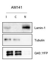
Several methods have been developed for the detection of insoluble protein aggregates including, for instance, dye binding assays (e.g., Thioflavin T, Congo red, NIAD-4) and electron microscopy. Filter retardation assay is a quick and sensitive method that detects and quantifies protein aggregates formed in vivo and in vitro, including polyglutamine (Scherzinger et al., 1997; Wanker et al., 1999), alpha-synuclein (Recasens et al., 2018), and amyloid-beta aggregates (Bieschke et al., 2009). In this assay, sodium dodecyl sulfate (SDS)-resistant protein aggregates are filtered and retained in a cellulose acetate membrane, while monomeric intermediate species are not captured. The protein aggregates retained in the membrane are subsequently detected by antibodies, which allows for their quantification.
This protocol describes a method to detect and quantify SDS-resistant polyglutamine aggregates in the nematode Caenorhabditis elegans and can be applied to investigate the aggregation of aggregation-prone proteins in vivo and in vitro.
Materials and Reagents
- 200 µl pipette tips (Greiner Bio-one, catalog number: 741065 )
- 1000 µl pipette tips (Greiner Bio-one, catalog number: 741045 )
- 15 ml conical tubes (SARSTEDT, catalog number: 62.554.502 )
- 2 ml Screw cap tubes (SARSTEDT, catalog number: 72.693.105 )
- 0.5 mm Glass beads (Carl Roth, catalog number: N030.1 )
- 11.3 x 7.7 cm Bio-Dot/Bio-Dot SF Filter Paper (Bio-Rad Laboratories, catalog number: 1620161 )
- Cellulose acetate membranes, 0.22 micron, Sphaero (Sterlitech, catalog number: CA023001 )
- 1.5 ml microfuge tube (Greiner Bio One International, catalog number: 616201 )
- Rubber seal
- Plastic wrap
- 50 ml conical tube (SARSTEDT, catalog number: 62.547.004 )
- Worms
- PierceTM BCA Protein Assay Kit (Thermo Fisher Scientific, catalog number: 23227 )
- Complete protease inhibitors (Roche Diagnostics, catalog number: 11697498001 )
- ECLTM Prime Western Blotting Detection (GE Healthcare, Amersham, catalog number: RPN2232 )
- Liquid nitrogen
- Triton X-100 (Sigma-Aldrich, catalog number: T8787 )
- DL-Dithiothreitol (DTT) (Sigma-Aldrich, catalog number: D0632 )
- 1 M Tris-HCl
- 5 M NaCl
- 10% SDS
- Milli-Q water
- Non-fat dry milk powder (Campina)
- Antibodies against green fluorescent protein (GFP)/yellow fluorescent protein (YFP) and tubulin (Table 1)
Table 1. List of primary and secondary antibodiesPrimary antibody Host Company/catalog number Dilution primary antibody Secondary antibody α-GFP Mouse TaKaRa Bio,Clontech Laboratories ( 632381 )1:5,000 α-tubulin Mouse Sigma-Aldrich ( T6074 ) 1:5,000 Anti-mouse (1:10,000)
Bio-Rad ( 1706516 ) - Sodium phosphate dibasic heptahydrate (Na2HPO4•7H2O) (Acros Organics, catalog number: 424380010 )
- Sodium chloride (NaCl) (Merck, catalog number: 1064041000 )
- Potassium chloride (KCl) (Fisher Chemicals, catalog number: 10010310 )
- 10x Phosphate buffered saline (PBS) (see Recipes)
- 1x PBS-Triton 0.1% (see Recipes)
- 1x PBS-Triton 0.05% (see Recipes)
- Filter trap assay (FTA) sample buffer (see Recipes)
- FTA wash buffer (see Recipes)
- 1 M dithiothreitol (DTT, see Recipes)
- Blocking solution (see Recipes)
Equipment
- P20 Pipetman (Gilson, catalog number: F123600 )
- P200 Pipetman (Gilson, catalog number: F123601 )
- P1000 Pipetman (Gilson, catalog number: F123602 )
- 30-300 μl multi-channel pipette (Eppendorf, catalog number: 3125000052 )
- 10 ml Serological glass pipettes (SARSTEDT, catalog number: 86.1254.001 )
- Centrifuge (Thermo Fisher Scientific, model: SL 40R , rotor: TX-750 )
- Bead beater (MP Biomedicals, model: FastPrep-24 )
- 48-well Bio-Dot Microfiltration System (Bio-Rad Laboratories, catalog number: 1703938 )
- Vacuum pump
- 20 °C incubator (LIEBHERR, model: WK 4126 )
- Vortex (Fisher scientific, model: ZX Wizard vortex, catalog number: 11746744 )
- Fuji Super RX-N 13x18 film (Fujifilm, catalog number: RX1318 )
- Autoclave
Software
- ImageJ (Open source: https://imagej.nih.gov/ij/)
- Microsoft Excel (Microsoft Corporation, Redmond, USA)
Procedure
文章信息
版权信息
© 2018 The Authors; exclusive licensee Bio-protocol LLC.
如何引用
Sin, O., Mata-Cabana, A., Seinstra, R. I. and Nollen, E. A. A. (2018). Filter Retardation Assay for Detecting and Quantifying Polyglutamine Aggregates Using Caenorhabditis elegans Lysates. Bio-protocol 8(19): e3042. DOI: 10.21769/BioProtoc.3042.
分类
生物化学 > 蛋白质 > 定量
您对这篇实验方法有问题吗?
在此处发布您的问题,我们将邀请本文作者来回答。同时,我们会将您的问题发布到Bio-protocol Exchange,以便寻求社区成员的帮助。