

Direct Visualization of the Multicopy Chromosomes in Cyanobacterium Synechococcus elongatus PCC 7942
蓝藻Synechococcus elongatus PCC 7942中多拷贝染色体的直接可视化观察


发布: 2018年08月05日第8卷第15期 DOI: 10.21769/BioProtoc.2958 浏览次数: 6967
评审: Anonymous reviewer(s)
参见作者原研究论文
The authors used this protocol in:

Jan 2018
Advertisement


Abstract
Cyanobacteria are prokaryotic organisms that carry out oxygenic photosynthesis. The fresh water cyanobacterium Synechococcus elongatus PCC 7942 is a model organism for the study of photosynthesis and gene regulation, and for biotechnological applications. Besides several freshwater cyanobacteria, S. elongatus 7942 also contains multiple chromosomal copies per cell at all stages of its cell cycle. Here, we describe a method for the direct visualization of multicopy chromosomes in S. elongatus 7942 by fluorescence in situ hybridization (FISH).
Keywords: Cyanobacteria (蓝藻)Background
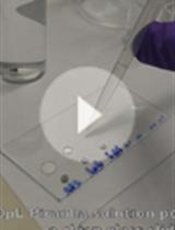
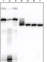
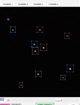
Cyanobacteria are prokaryotic microorganisms that utilize an oxygen-producing photosynthetic system similar to that of chloroplasts in higher plants. Whereas bacteria such as Escherichia coli and Bacillus subtilis harbor a single circular chromosome, several species of freshwater cyanobacteria have multiple circular chromosomes per cell (Mann and Carr, 1974; Labarre et al., 1989; Binder and Chisholm, 1995). The freshwater cyanobacterium Synechococcus elongatus PCC 7942 (hereafter referred to as S. elongatus 7942) carries 3-8 chromosomal copies per cell (Griese et al., 2011; Watanabe et al., 2012; Watanabe et al., 2015). A fluorescence reporter-operator system has been established to acquire images of the multicopy chromosome of S. elongatus 7942 (Chen et al., 2012; Jain et al., 2012). Although this system enables live-cell imaging, it requires complex genetic constructs for labeling individual chromosomes, and, overall, this type of system is unstable over an extended period of cultivation. Thus, it is necessary to examine the chromosomal distribution in S. elongatus 7942 cells that have a simple genetic background. We recently established a fluorescence in situ hybridization (FISH) method for visualizing the oriC region and putative terC region of the multicopy chromosome in S. elongatus 7942 (Watanabe et al., 2018). A DNA probe, which covers the oriC or terC region, was synthesized and used for the FISH analysis. Labeled chromosomes in S. elongatus 7942 cells can be observed by fluorescence microscopy. Here, we describe a step-by-step protocol for the FISH method.
Materials and Reagents
- Pipette tips
200 μl (FUKAEKASEI and WATSON, catalog number: 110-705C )
1,000 μl (FUKAEKASEI and WATSON, catalog number: 110-706C ) - PCR-tubes and strips
Tube (FUKAEKASEI and WATSON, catalog number: 137-333C )
Cap (FUKAEKASEI and WATSON, catalog number: 137-432C ) - Disposable plastic dish (for BG-11 plate) (KANTO KAGAKU, model: CSPD90-15, catalog number: 96930-01 )
- 93 ml test tubes (for culturing) (MonotaRO, IWAKI, catalog number: 09184061)
Manufacturer: IWAKI, catalog number: TEST30NP . - Poly-lysine coated glass slides (Matsunami Glass, catalog number: S7441 )
- Cover glass (MonotaRO, catalog number: CT18189 )
- Tube for sonication (microtube AFA Fiber Pre-Slit Snap-Cap) (Covaris, catalog number: 520077 )
- Aluminum foil
- Synechococcus elongatus PCC 7942 strain (Pasteur culture collection)
- Genomic DNA of S. elongatus 7942 (10-50 ng for each PCR reaction)
- ExTaq PCR Kit (TaKaRa Bio, catalog number: RR001A )
- Fluorescence-12-dUTP (Roche Diagnostics, catalog number: 11427857910 )
- Random primed DNA labeling kit (Roche Diagnostics, catalog number: 11004760001 )
- 99.5% ethanol (KANTO KAGAKU, catalog number: 14032-08 )
- 3 M NaOAc solution (pH 5.2) (KANTO KAGAKU, catalog number: 37092-00 )
- Glycogen (20 ng/ml) (Roche Diagnostics, catalog number: 10901393001 )
- Triton X-100 (NACALAI TESQUE, catalog number: 35501-02 )
- Formamide (KANTO KAGAKU, catalog number: 16062-00 )
- Paraformaldehyde (KANTO KAGAKU, catalog number: 32034-12 )
- Dimethyl sulfoxide (KANTO KAGAKU, catalog number: 10378-00 )
- Sodium hydroxide (KANTO KAGAKU, catalog number: 37184-00 )
- Methanol (KANTO KAGAKU, catalog number: 25183-00 )
- PBS powder (Sigma-Aldrich, catalog number: P3813 )
- Lysozyme (Wako Pure Chemical Industries, catalog number: 122-02673 )
- Tris-HCl (pH 7.5) (Sigma-Aldrich, catalog number: T3253-250G )
- EDTA (NACALAI TESQUE, catalog number: 15111-45 )
- Sodium chloride (KANTO KAGAKU, catalog number: 37144-00 )
- Trisodium citrate dihydrate (KANTO KAGAKU, catalog number: 37150-00 )
- Glycerol (KANTO KAGAKU, catalog number: 17029-00 )
- DAPI (Sigma-Aldrich, catalog number: D9564-10MG )
- Salmon sperm DNA (Roche Diagnostics, catalog number: 11467140001 )
- Three sets of PCR primers for visualizing oriC or terC (Eurofins Genomics, Table 1)
Table 1. List of primers used for the preparation of FISH probes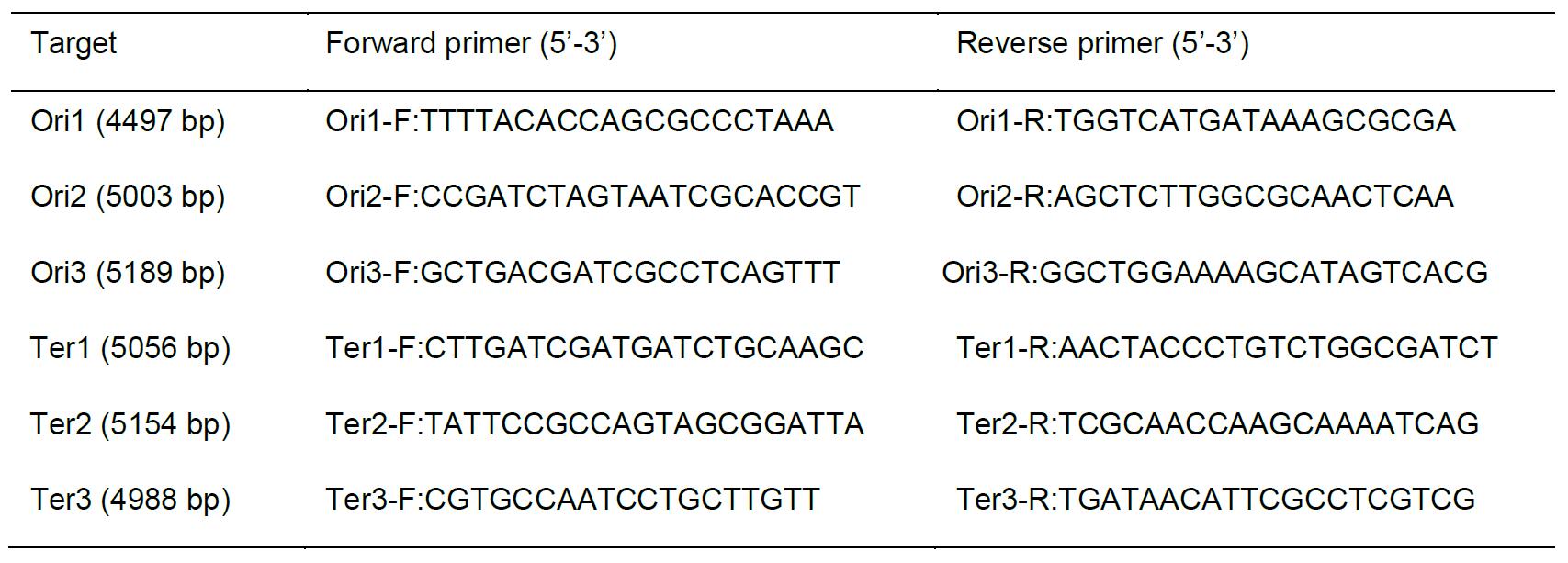
- BG-11 liquid medium (pH 8.2) (Castenholz, 1988)
- BG-11 plates (pH 8.2) (Castenholz, 1988)
- Hybridization solution (see Recipes)
- Fixation solution (see Recipes)
- PBS buffer (see Recipes)
- Lysozyme solution (see Recipes)
- 20x Standard Saline Citrate (SSC) (see Recipes)
- Mounting solution (see Recipes)
Equipment
- Pipettes
P20 (Gilson, catalog number: F123600 )
P200 (Gilson, catalog number: F123601 )
P1000 (Gilson, catalog number: F123602 ) - Thermal cycler (Thermo Fisher Scientific, Applied Biosystems, model: Veriti )
- Microtube centrifuge (TOMY SEIKO, model: MX-107 )
- Covaris S-2 sonicator (Covaris, Woburn, MA, USA)
- Plant growth chamber (TOMY SEIKO, model: CLE-405 )
- UV/Vis spectrophotometer (Shimadzu, model: UV-1800 )
- Pharmaceutical refrigerator (Panasonic, model: MPR-3120CN-PJ )
- Glass vat for staining (AS ONE, models: 1-4398-01 and 1-4398-11 )
- Forma direct heat CO2 Incubator (Thermo Fisher Scientific, model: FormaTM 310 , catalog number: 320)
- Fluorescence microscope (Olympus, models: BX53 and DP71 ; filters: U-FUW for DAPI and U-FGW for chlorophyll, objective: UPLSAPO 100XOPH)
- All-in-one fluorescence microscope (Olympus, model: FSX100 , filters: U-MNUA2 for DAPI, U-MWIBA3 for FISH signal, and U-MWIG3 for chlorophyll, objective: UPLSAPO , super apochromat)
Note: This system is necessary to obtain clear FISH images.
Software
- Camera software for DP71 (Olympus)
- Software for FSX 100 (Olympus, FSX-BSW)
- Adobe Photoshop Elements 11 (Adobe Photoshop, CC)
Procedure
文章信息
版权信息
© 2018 The Authors; exclusive licensee Bio-protocol LLC.
如何引用
Ohbayashi, R., Yoshikawa, H. and Watanabe, S. (2018). Direct Visualization of the Multicopy Chromosomes in Cyanobacterium Synechococcus elongatus PCC 7942. Bio-protocol 8(15): e2958. DOI: 10.21769/BioProtoc.2958.
分类
微生物学 > 微生物细胞生物学 > 细胞染色
分子生物学 > DNA > DNA 标记
细胞生物学 > 细胞成像 > 荧光
您对这篇实验方法有问题吗?
在此处发布您的问题,我们将邀请本文作者来回答。同时,我们会将您的问题发布到Bio-protocol Exchange,以便寻求社区成员的帮助。