

HCV Reporter System (Viral Infection-Activated Split-Intein-Mediated Reporter System) for Testing Virus Cell-to-cell Transmission ex-vivo
用于测试病毒细胞间传递的HCV报告系统(病毒感染-激活的断裂内含肽介导的报告系统)
发布: 2018年08月05日第8卷第15期 DOI: 10.21769/BioProtoc.2949 浏览次数: 6449
评审: David PaulJose Antonio Reyes-DariasAnonymous reviewer(s)
参见作者原研究论文
The authors used this protocol in:

Jan 2017
Advertisement


Abstract
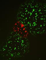
Hepatitis C virus (HCV) spread involves two distinct entry pathways: cell-free transmission and cell-to-cell transmission. Cell-to-cell transmission is not only an efficient way for viruses to spread but also an effective method for escaping neutralizing antibodies. We adapted the viral infection-activated split-intein-mediated reporter system (VISI) and developed a straightforward model for Live-cell monitoring of HCV cell-to-cell transmission ex-vivo: co-culture of HCV infected donor cells (red signal) with uninfected recipient cells (green signal) and elimination of the cell-free transmission by adding potent neutralizing antibody AR3A in the supernatant. With this model, the efficiency of cell-to-cell transmission can be evaluated by counting the number of foci designated by the green signal of recipient cells.
Keywords: HCV (HCV)Background
Accumulating evidence support that viruses can use different routes of spread in infected tissues (Sattentau, 2008; Zhong et al., 2013). For HCV transmission, both cell-free transmission and cell-to-cell transmission can mediate virus transfer between hepatocytes. While cell-free transmission initiates HCV infection, cell-cell transmission is thought to transfer HCV to adjacent hepatocytes directly. It provides an excellent way to resist the neutralizing antibodies and contribute to the viral persistence (Brimacombe et al., 2011; Xiao et al., 2014). Previous articles also proved some host factors which contributed to cell-cell transmission, such as scavenger receptor BI (SR-BI), CD81, tight junction proteins claudin-1 (CLDN1), Occludin (OCLN), epidermal growth factor receptor (EGFR) (Witteveldt et al., 2009; Catanese et al., 2013; Zona et al., 2013). But the exact mechanisms of this process still need to explore. We optimized a viral infection-activated split-intein-mediated reporter system (VISI) for live-cell visualization of HCV infection (Zhao et al., 2017). Based on the study in Huh7.5.1 cell line using a technique of split GFP/RFP reconstitution by intein protein splicing (Figure 1A), it showed that VISI system is a very sensitive and low background system. With this system, we can clearly visualize the HCV infected cell by its nuclear fluorescence signal. In addition, combining VISI-GFP and VISI-mCherry cells, we can further monitor HCV cell-to-cell transmission in the presence of potent neutralizing antibody AR3A.
Materials and Reagents
- For cell culture materials
- Sterile 100 mm polystyrene Petri dish (Thermo Fisher Scientific, catalog number: 172931 )
- Sterile flat-bottom 96-well plate (Thermo Fisher Scientific, catalog number: 167008 )
- Sterile 60 mm polystyrene Petri dish (Thermo Fisher Scientific, catalog number: 150288 )
- Sterile 6-well plate (Thermo Fisher Scientific, catalog number: 140675 )
- Sterile 50 ml Conical Centrifuge Tube (Thermo Fisher Scientific, catalog number: 339652 )
- Sterile 100 mm polystyrene Petri dish (Thermo Fisher Scientific, catalog number: 172931 )
- Plasmids and Cell lines
- Lentiviral vector:
pWPI-blasticidin-NLS-GFPn-INTEINn (Genbank: KY067203)
pWPI-puromycin-INTEINc-GFPc-NLS-IPS (Genbank: KY067204) (for construction of Huh7.5.1-VISI-GFP cell line)
pWPI-blasticidin-NLS-mCherry(n)-INTEINn (Genbank: KY067205)
pWPI-puromycin-INTEINc-mCherry(c)-NLS-IPS (Genbank: KY067206) (for construction of Huh7.5.1-VISI-mCherry cell line) - Two auxiliary plasmids:
psPAX2 (the HIV-1 packaging plasmid)
pMD2.G (a vesicular stomatitis virus glycoprotein [SV-G] expression vector)
- 293T cell (ATCC, catalog number: CRL-3216 )
- Huh7.5.1 cell (Human hepatocyte-derived cell line Huh7.5.1 is kindly provided by professor F. Chisari)
- Lentiviral vector:
- HCV virus strains
- HCV JC1 (GenBank: JF343782.1)
- Subgenomic JFH1 (sgJFH1) (GenBank: AB114136.1)
- HCV JC1 (GenBank: JF343782.1)
- For cell culture medium
- 1x phosphate-buffered saline (PBS) (Thermo Fisher Scientific, GibcoTM, catalog number: 10010049 )
- 0.25% Trypsin (Thermo Fisher Scientific, GibcoTM, catalog number: 25200072 )
- DMEM (Thermo Fisher Scientific, GibcoTM, catalog number: C11965500CP )
- Fetal bovine serum (Thermo Fisher Scientific, GibcoTM, catalog number: 10099141 )
- 100x Nonessential amino acids (Thermo Fisher Scientific, GibcoTM, catalog number: 11140050 )
- 100x Penicillin/streptomycin (Thermo Fisher Scientific, GibcoTM, catalog number: 15140122 )
- Complete DMEM (see Recipes)
- 1x phosphate-buffered saline (PBS) (Thermo Fisher Scientific, GibcoTM, catalog number: 10010049 )
- For transcription in vitro
- MEGAscript® T7 Transcription Kit (Thermo Fisher Scientific, catalog number: AM1333 )
- MEGAscript® T7 Transcription Kit (Thermo Fisher Scientific, catalog number: AM1333 )
- For electroporation buffer
- ATP (Thermo Fisher Scientific, catalog number: R0441 )
- L-Glutathione (Sigma-Aldrich, catalog number: V900456-25G )
- Potassium chloride (KCl) (Sinopharm Chemical Reagent, catalog number: 10016318 )
- Calcium chloride (CaCl2) (Shanghai Experiment Reagent, catalog number: 117600 )
- Dipotassium hydrogen phosphate (K2HPO4) (Shanghai Experiment Reagent, catalog number: 168120 )
- Potassium phosphate monobasic (KH2PO4) (Shanghai Experiment Reagent, catalog number: 175650 )
- HEPES (Sigma-Aldrich, catalog number: H4034-25G )
- EGTA (Sangon Biotech, catalog number: E0732-50G )
- Magnesium chloride (MgCl2) (Sinopharm Chemical Reagent, catalog number: 10012818 )
- Cytomix buffer (see Recipes)
- ATP (Thermo Fisher Scientific, catalog number: R0441 )
- For Calcium Phosphate (CaPO4) transfection buffer
- HEPES (Sigma-Aldrich, catalog number: H4034-25G )
- Sodium chloride (NaCl) (Sinopharm Chemical Reagent, catalog number: 10019318 )
- Disodium hydrogen phosphate (Na2HPO4·12H2O) (Shanghai Experiment Reagent, catalog number: 174710 )
- Calcium chloride (CaCl2) (Shanghai Experiment Reagent, catalog number: 117600 )
- Calcium Phosphate (CaPO4) transfection buffer (see Recipes)
- HEPES (Sigma-Aldrich, catalog number: H4034-25G )
- HCV-neutralizing antibody
- Antibody AR3A
Note: AR3A is kindly provided by professor M. Law.
- Antibody AR3A
- For cell line selecting
- Puromycin (Sigma-Aldrich, catalog number: P8833-25MG )
- Blasticidin (Thermo Fisher Scientific, GibcoTM, catalog number: R21001 )
- Puromycin (Sigma-Aldrich, catalog number: P8833-25MG )
Equipment
- Electroporator (Bio-Rad Laboratories, model: Gene Pulser XcellTM )
- Electroporation cuvette (Bio-Rad Laboratories, Gene Pulser cuvette, 0.4 cm)
- Fluorescence microscope (Olympus, model: IX53 )
Software
- GraphPad Prism (GraphPad Software, https://www.graphpad.com/)
Procedure
文章信息
版权信息
© 2018 The Authors; exclusive licensee Bio-protocol LLC.
如何引用
Zhao, F., Zhao, T., Deng, L., Lv, D., Zhang, X., Pan, X., Xu, J. and Long, G. (2018). HCV Reporter System (Viral Infection-Activated Split-Intein-Mediated Reporter System) for Testing Virus Cell-to-cell Transmission ex-vivo. Bio-protocol 8(15): e2949. DOI: 10.21769/BioProtoc.2949.
分类
微生物学 > 体内实验模型 > 病毒
细胞生物学 > 细胞成像 > 活细胞成像
您对这篇实验方法有问题吗?
在此处发布您的问题,我们将邀请本文作者来回答。同时,我们会将您的问题发布到Bio-protocol Exchange,以便寻求社区成员的帮助。