

3D Culture Protocol for Testing Gene Knockdown Efficiency and Cell Line Derivation
用于检测基因敲减效率和细胞系建立3D培养方法

发布: 2018年06月05日第8卷第11期 DOI: 10.21769/BioProtoc.2874 浏览次数: 10913
评审: Chiara AmbrogioMauro Sbroggio'Enrico Patrucco
参见作者原研究论文
The authors used this protocol in:
Apr 2017


Abstract
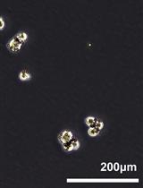
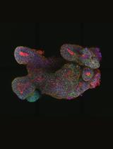
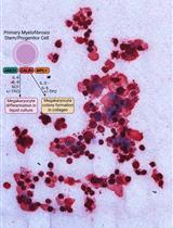
Traditional 2D cell cultures with cells grown as monolayers on solid surface still represent the standard method in cancer research for drug testing. Cells grown in 2D cultures, however, lack relevant cell-matrix and cell-cell interactions and ignore the true three-dimensional anatomy of solid tumors. Cells cultured in 2D can also undergo cytoskeletal rearrangements and acquire artificial polarity associated with aberrant gene expression (Edmondson et al., 2014). 3D culture systems that better mimic the in vivo situation have been developed recently. 3D in vitro cancer models (tumorspheres) for studying cancer stem cells have gained increased popularity in the field (Weiswald et al., 2015). Systems that use matrix-embedded or encapsulated spheroids, spheroids cultured in hanging drops, magnetic levitation systems or 3D printing methods are already being widely used in research and for novel drug screening. In this article, we describe a detailed protocol for testing the effect of shRNA-mediated gene silencing on tumorsphere formation and growth. This approach allows researchers to test the impact of gene knockdown on the growth of tumor initiating cells. As verified by our lab, the protocol can be also used for isolation of 3D cancer cell lines directly from tumor tissues.
Keywords: 3D culture (三维培养)Background
3D in vitro cancer cell models represent a bridge experimental method between cell lines and tumors grown in vivo (Pampaloni et al., 2007; Weiswald et al., 2015). 3D characters of solid tumors with heterogeneous access to nutrients or oxygen can only be effectively mimicked by 3D culture systems. In recent years, protocols for tumorsphere culture gained lot of interest. A tumorsphere can be described as a solid, spherical object created from a single progenitor or stem cell. For tumorspheres formation assays, cells are seeded and grown in serum-free media in ultra-low attachment plates (non-adherent conditions), which allows enrichment of cancer cells with stem/progenitor properties (Johnson et al., 2013). Tumorspheres generated from freshly isolated tumor tissue are of special interest in the field because cells from established cell lines typically differ from the primary tumor due to mutations and abnormalities gained during multiple rounds of in vitro passaging. Hereby, we present an optimized protocol for 3D culture-based primary tumor cell isolation and the use of 3D culture to assess the effect of gene silencing on the growth of tumor-initiation cells.
Materials and Reagents
- Eppendorf tube
- Pipette tips
- Vials
- Plastic bottles
- 50 ml Falcon tube
- Petri dishes with clear lid (Fisher Scientific, Fisherbrand, catalog number: FB0875712 )
- 6-well plates, Corning Costar Ultra-Low Attachment (Corning, catalog number: 3471 )
- 15 ml Falcon tubes (Corning, Falcon®, catalog number: 352196 )
- 70 µm cell filter (cell strainer - Corning, Falcon®, catalog number: 352350 )
- Ice
- Plastic pipette
- 24-well plates, Corning Costar Ultra-Low Attachment (Corning, catalog number: 3473 )
- 6-well plates (Corning, catalog number: 3506 )
- Serological pipettes
- Surgical razor blades (FisherBrand High Precision # 22 Style Scalpel Blade, Fisher Scientific, FisherbrandTM, catalog number: 12-000-161 )
- Sterile pipets (10 ml) (FisherBrand Sterile Disposable Standard Serological Pipets, Fisher Scientific, FisherbrandTM, catalog number: 13-678-14A )
- Cryovials (General Long-Term Storage Cryogenic Tubes 1 ml, Thermo Fisher Scientific, Thermo ScientificTM, catalog number: 5000-1012 )
- (optional) Mission pLKO.1-puro-CMV-TurboGFP Positive control Transduction Particles (Sigma-Aldrich, catalog number: SHC003V )
- EDTA (Thermo Fisher Scientific, InvitrogenTM, catalog number: 15575-020 )
- PBS pH 7.4 (Thermo Fisher Scientific, GibcoTM, catalog number: 10010023 )
- Trypsin EDTA (0.05%) (Thermo Fisher Scientific, GibcoTM, catalog number: 25300054 )
- SureEntry transduction reagent (QIAGEN, catalog number: 336921 )
- Clorox Bleach (Veritiv, catalog number: 30966 )
- Heparin Sodium Salt (Sigma-Aldrich, catalog number: H3149-10KU )
- DMEM/F12, Glutamax (Thermo Fisher Scientific, GibcoTM, catalog number: 10565018 )
- Penicillin/Streptomycin (Thermo Fisher Scientific, GibcoTM, catalog number: 15070063 )
- Fetal bovine serum (Thermo Fisher Scientific, GibcoTM, catalog number: 26140079 )
- B27 supplement 50x (serum free) (Thermo Fisher Scientific, GibcoTM, catalog number: 17504044 )
- bFGF – human Fibroblasts Growth Factor 147 basic (animal free) (Gemini Bio-Products, catalog number: 300-805P )
- EGF– Epidermal Growth Factor (human) (Gemini Bio-Products, catalog number: 300-110P )
- ROCK inhibitor (Y-27632) (STEMCELL Technologies, catalog number: 72304 )
- Heparin solution (STEMCELL Technologies, catalog number: 07980 )
- BD Matrigel Matrix Growth Factor Reduced (BD Biosciences, catalog number: 356230 ) or Cultrex® 3-D Culture MatrixTM Reduced Growth Factor Basement Membrane Extract (Trevigen, catalog number: 3445-001-01 )
Notes:- Aliquot Matrigel or Cultrex Matrix into single-use aliquots in a sterile hood into sterile Eppendorf tubes. We recommend 0.5 ml of Matrigel per Eppendorf tube. When combined with 1.5 ml of ice-cold media, this amount is sufficient for 3D culture using one well in a six-well plate.
- Use chilled pipettes and have Eppendorf tubes on ice during procedure.
- Store at -80 °C. Thaw on ice or in the refrigerator overnight before use.
- Aliquot Matrigel or Cultrex Matrix into single-use aliquots in a sterile hood into sterile Eppendorf tubes. We recommend 0.5 ml of Matrigel per Eppendorf tube. When combined with 1.5 ml of ice-cold media, this amount is sufficient for 3D culture using one well in a six-well plate.
- Hibernation medium (HibernateTM-A, Thermo Fisher Scientific, GibcoTM, catalog number: A1247501 )
- Collagenase IV (Sigma-Aldrich, catalog number: C8051-100MG or similar)
- Trypan Blue (TC-10, Bio-Rad Laboratories, catalog number: 1450013 )
- Antibiotic-Antimycotic (100x) (Thermo Fisher Scientific, GibcoTM, catalog number: 15240096 )
- Dispase (1 U/ml, STEMCELL Technologies, catalog number: 07923 )
- EDTA (Thermo Fisher Scientific, InvitrogenTM, catalog number: 15575020 )
- Accutase (STEMCELL Technologies, catalog number: 07920 )
- DMSO (Sigma-Aldrich, catalog number: D2438-50ML )
- Ethanol (70% solution, Fisher Scientific, Fisher BioReagentsTM, catalog number: BP8201500 )
- Culture medium (see Recipes)
- CSC medium (see Recipes)
Equipment
- Centrifuge (temperature controlled, VWR® benchtop general purpose centrifuge) (VWR, catalog number: 10830-764 )
- Pipettes
- Forceps
- CO2 Cell incubator (BINDER, model: CB 160 )
- Biological Safety Level (BSL-2) laminar flow hood (Esco, EscoTechnologies, USA)
- TC20 Automated cell counter (Bio-Rad)
- Mr. Frosty Freezing Container (Thermo Fisher Scientific, Thermo ScientificTM, catalog number: 5100-0001 )
- FormaTM 7000 series Ultra Low Temperature Freezers (Thermo Fisher Scientific, Thermo ScientificTM, model: TFM#902 Series )
- Liquid nitrogen storage tank
- Nikon TE-2000E D-Eclipse Csi confocal microscope running Nikon Elements software (Nikon, model: Eclipse TE2000-E )
Software
- Nikon microscope operating software
- ImageJ or similar image processing software (https://imagej.nih.gov/ij/)
- GraphPad Prism 6.0 software from GraphPad Software, USA
(https://www.graphpad.com/scientific-software/prism/) - R software (R Core Team, 2015 R: A language and environment for statistical computing)
Procedure
文章信息
版权信息
© 2018 The Authors; exclusive licensee Bio-protocol LLC.
如何引用
Strnadel, J., Woo, S. M., Choi, S., Wang, H., Grendar, M. and Fujimura, K. (2018). 3D Culture Protocol for Testing Gene Knockdown Efficiency and Cell Line Derivation. Bio-protocol 8(11): e2874. DOI: 10.21769/BioProtoc.2874.
分类
癌症生物学 > 癌症干细胞 > 细胞生物学试验 > 细胞分离和培养
细胞生物学 > 细胞信号传导 > 胞内信号传导
您对这篇实验方法有问题吗?
在此处发布您的问题,我们将邀请本文作者来回答。同时,我们会将您的问题发布到Bio-protocol Exchange,以便寻求社区成员的帮助。