

Intracellular and Mitochondrial Reactive Oxygen Species Measurement in Primary Cultured Neurons
原代培养的神经元细胞和线粒体中活性氧的测定
发布: 2018年06月05日第8卷第11期 DOI: 10.21769/BioProtoc.2871 浏览次数: 11901
评审: Xi FengWelsch Charles JeremyAnonymous reviewer(s)
参见作者原研究论文
The authors used this protocol in:

May 2017
Advertisement


Abstract
Reactive oxygen species (ROS) are chemically reactive oxygen containing molecules. ROS consist of radical oxygen species including superoxide anion (O2•−) and hydroxyl radical (•OH) and non-radical oxygen species such as hydrogen peroxide (H2O2), singlet oxygen (O2). ROS are generated by mitochondrial oxidative phosphorylation, environmental stresses including UV or heat exposure, and cellular responses to xenobiotics (Ray et al., 2012). Excessive ROS production over cellular antioxidant capacity induces oxidative stress which results in harmful effects such as cell and tissue damage. Sufficient evidence suggests that oxidative stresses are involved in cancers, cardiovascular disease, and neurodegenerative diseases including Alzheimer’s disease and Parkinson disease (Waris and Ahsan, 2006). Though excessive level of ROS triggers detrimental effects, ROS also have been implicated to regulate cellular processes. Since ROS function is context dependent, measurement of ROS level is important to understand cellular processes (Finkel, 2011). This protocol describes how to detect intracellular and mitochondrial ROS in live cells using popular chemical fluorescent dyes.
Keywords: Reactive oxygen species (ROS) (活性氧族(ROS))Background
ROS are important to maintain homeostasis in our bodies (Brieger et al., 2012). Many diseases such as cancer, neurodegenerative disease, cardiovascular disease, and diabetics are associated with ROS (Datta et al., 2000). DNA damage caused by ROS is a major cause of accelerating carcinogenesis process, and therapeutic agents targeting ROS have been actively developed (Trachootham et al., 2009). In circulatory system, abnormal oxidative stress increases the production of ROS, leading to various cardiovascular diseases (Forstermann, 2008). Signaling related to diabetes is sensitive to ROS, and these signaling abnormalities induced by abnormal levels ROS cause diabetes complications (Baek et al., 2017). Controlling the ROS levels in the brain is one of the most important activities because abnormal levels of ROS can cause diverse brain diseases. Amyloid beta, known as an important factor in Alzheimer’s disease, causes excessive ROS generation in the brain, neuronal damage (Singh et al., 2011), and eventually dementia (Polidori, 2004). Activated microglia produced by ROS which secretes a variety of cytokines result in neuronal death (Heneka et al., 2014).
ROS are generated by small part of oxygen consumed in mitochondria. A principal species of ROS produced in mitochondria is superoxide anion and it is the byproduct of the electron transport chain (Batandier et al., 2002). In order to detect superoxide in mitochondria, MitoSOX red, a mitochondria superoxide indicator, is used. Due to the positive charge on triphenylphosphonium group, MitoSOX red can effectively penetrate phospholipid bilayer, and accumulate into the matrix of mitochondria. Furthermore, hydroethidine of MitoSOX red allows researchers to discriminate the fluorescent signal generated by superoxide-mediated oxidative products from other non-specific signals (Robinson et al., 2006; Baek et al., 2017).
CM-H2DCFDA is a chloromethyl derivative of H2DCFDA (2',7'-dichlorodihydrofluorescein diacetate), a fluorogenic dye that measures hydroxyl, peroxyl and other ROS activity within the cell and can be used to detect the intracellular formation of ROS (Kirkland et al., 2007). Once the fluorescent probe of CM-H2DCFDA permeates cell membrane, intracellular esterases hydrolyze its acetyl groups and it can be retained in the cell. CM-H2DCFDA is more sensitive to oxidation by H2O2 than superoxide (O2•−) (Fowler et al., 2017). CM-H2DCFDA is widely used in physiological and pathophysiological studies including virus infection (Nykky et al., 2014), cancer (Khatri et al., 2015; Liu et al., 2017), and neurodegenerative diseases (Ng et al., 2014). Using CM-H2DCFDA, we can detect intracellular ROS level by flow cytometry/fluorescence measurement and the localization of ROS producing organelle with confocal microscopy (Forkink et al., 2010).
Materials and Reagents
- Glass bottom cell culture dish type 35 mm and dimension 20 mm (Nest Scientific, catalog number: 801001 )
- Cover glasses thickness No. 1 circular size 18 mm Ø (MARIENFELD, catalog number: 0111580 )
- Petri dish, 100 mm Polysterene aseptic non-tissue culture treated (SPL Life Sciences, catalog number: 10095 )
- 15 ml conical tube (SPL Life Sciences, catalog number: 50015 )
- 10 ml Serological pipettes (SPL Life Sciences, catalog number: 91010 )
- 50 ml conical tube (SPL Life Sciences, catalog number: 50050 )
- Cell strainer 70 μm (Corning, Falcon®, catalog number: 352350 )
- Pregnant female Sprague Dawley rats (E17-E18 days gestation, Orient Korea)
- Poly-D-lysine hydrobromide (Sigma-Aldrich, catalog number: P6407-5mg )
- Phosphate buffered saline powder, pH 7.4, for preparing 1 L solutions (Sigma-Aldrich, catalog number: P3813 )
- CM-H2DCFDA (Thermo Fisher Scientific, InvitrogenTM, catalog number: C6827 )
- Dimethyl Sulfoxide(DMSO) (Merck, catalog number: 317275 )
- MitoSOXTM Red Mitochondrial Superoxide Indicator, for live-cell imaging (Thermo Fisher Scientific, InvitrogenTM, catalog number: M36008 )
- Phosphate buffered saline (PBS) powder, pH 7.4, for preparing 1 L solutions, suitable for cell culture(Sigma-Aldrich, catalog number: P3813 )
- Trypsin (2.5%), no phenol red (Thermo Fisher Scientific, GibcoTM, catalog number: 15090046 )
- Fetal bovine serum (FBS) (Thermo Fisher Scientific, GibcoTM, catalog number: 10082147 )
- Neurobasal Medium® (Thermo Fisher Scientific, GibcoTM, catalog number: 21103049 )
- B-27TM Supplement (50x), serum free (Thermo Fisher Scientific, GibcoTM, catalog number: 17504044 )
- DMEM High Glucose (4.5 g/L), with L-Glutamine, with Sodium Pyruvate (Capricorn Scientific, catalog number: DMEM-HPA )
- Penicillin/Streptomycin (100x) (PS) (Capricorn Scientific, catalog number: PS-B )
- Amyloid beta peptide 1-42 Human (ANYGEN, catalog number: AGP-8338 )
- CM-H2DCFDA solution (see Recipes)
- MitoSOXTM Red solution (see Recipes)
- Poly-D-lysine hydrobomide solution (see Recipes)
- Prep medium (see Recipes)
- Culture medium (see Recipes)
- Maintain culture medium (see Recipes)
Equipment
- Haemacytometers (MARIENFELD, catalog number: 0630010 )
- Original Portable Pipet-Aid® Pipette Controller (Drummond Scientific, catalog number: 4-000-100 )
- Dressing Scissors (Surgimax Instruments, catalog number: 85-112-12 ) (Figure 1A 1)
- Dissecting Scissors (Surgimax Instruments, catalog number: 85-127-10 ) (Figure 1A 2)
- Dissecting Scissors (Surgimax Instruments, catalog number: 63-175-11 ) (Figure 1A 3)
- Spring Dressing Forceps Sharp (Surgimax Instruments, catalog number: 85-076-11 ) (Figure 1A 4)
- Spring Dressing Forceps Blunt (Surgimax Instruments, catalog number: 85-073-15 ) (Figure 1A 5)
- Multi Purpose Forceps Pointed (Surgimax Instruments, catalog number: 05-177-11 ) (Figure 1A 6)
- Clean bench (HANBAEK Scientific Technology, catalog number: HB-402 )
- Cell culture CO2 incubator (ARA, catalog number: APR150 )
- Water-bath (Grant Instruments, JB Academy, catalog number: JBA18 )
- Centrifuge (Hanil Scientific, catalog number: Combi 514R )
- Confocal microscope with live cell imaging system (Carl Zeiss, model: LSM700 ) (Figures 1B and 1C)
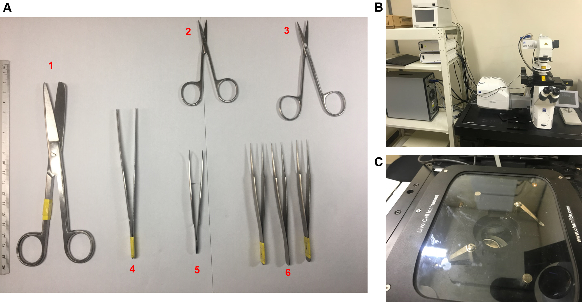
Figure 1. Equipment for the experiment. A. Surgery instruments; B. Confocal microscope (LSM700) with live cell imaging system; C. Live cell chamber.
Software
- For measure
- ZEN black version (ZEISS confocal microscope LSM700 software)
Note: This is default program provided with ZEISS confocal microscope.
- ZEN black version (ZEISS confocal microscope LSM700 software)
- For analysis
- ZEN Blue edition (ZEISS confocal microscope LSM700 software; SR-DIP software for ZEN blue edition)
- ImageJ (ImageJ is an open source image processing program)
- ZEN Blue edition (ZEISS confocal microscope LSM700 software; SR-DIP software for ZEN blue edition)
Procedure
文章信息
版权信息
© 2018 The Authors; exclusive licensee Bio-protocol LLC.
如何引用
Readers should cite both the Bio-protocol article and the original research article where this protocol was used:
- Baek, S. H., Cho, Y., Lee, J., Choi, B. Y., Choi, Y., Park, J. S., Kim, H., Sul, J., Kim, E., Park, J. H. and Jo, D. (2018). Intracellular and Mitochondrial Reactive Oxygen Species Measurement in Primary Cultured Neurons. Bio-protocol 8(11): e2871. DOI: 10.21769/BioProtoc.2871.
- Baek, S. H., Park, S. J., Jeong, J. I., Kim, S. H., Han, J., Kyung, J. W., Baik, S. H., Choi, Y., Choi, B. Y., Park, J. S., Bahn, G., Shin, J. H., Jo, D. S., Lee, J. Y., Jang, C. G., Arumugam, T. V., Kim, J., Han, J. W., Koh, J. Y., Cho, D. H. and Jo, D. G. (2017). Inhibition of Drp1 ameliorates synaptic depression, Aβ deposition, and cognitive impairment in an Alzheimer's disease model. J Neurosci 37(20): 5099-5110.
分类
神经科学 > 细胞机理 > 胞内信号传导
神经科学 > 神经系统疾病 > 细胞机制
细胞生物学 > 细胞成像 > 共聚焦显微镜
您对这篇实验方法有问题吗?
在此处发布您的问题,我们将邀请本文作者来回答。同时,我们会将您的问题发布到Bio-protocol Exchange,以便寻求社区成员的帮助。