

3D Co-culture System of Tumor-associated Macrophages and Ovarian Cancer Cells
肿瘤相关巨噬细胞和卵巢癌细胞的3D共培养系统

发布: 2018年04月20日第8卷第8期 DOI: 10.21769/BioProtoc.2815 浏览次数: 12191
评审: Jia LiLucíola Silva BarcelosAnonymous reviewer(s)
参见作者原研究论文
The authors used this protocol in:
Nov 2016
Advertisement


Abstract
Ovarian cancer is fairly unique in that ovarian carcinoma cells can detach and spread directly through peritoneal cavity. It has been unclear, however, how detached cancer cells survive in the peritoneum and form spheroid structure. We have recently reported that there is a strong correlation between Tumor-associated macrophages (TAMs)-associated spheroid and clinical pathology of ovarian cancer, and that TAMs promote spheroid formation and tumor growth at early stages of transcoelomic metastasis in orthotopic mouse models. We have established an in vitro spheroid formation assay using a 3D co-culture system in which mouse GFP+F4/80+CD206+ TAMs isolated from spheroids of ovarian cancer-bearing donor tomatolysM-cre mice were mixed with ID8 cells (TAM:ID8 at a ratio of 1:10) in medium containing 2% Matrigel and seeded onto the 24-well plate precoated with Matrigel. As transcoelomic metastasis is also associated with many other cancers such as pancreatic and colon cancers, TAM-mediated spheroid formation assay would provide a useful approach to define the molecular mechanism and therapeutic targets for ovarian cancer and other transcoelomic metastasis cancers.
Keywords: Ovarian cancer (卵巢癌)Background
Ovarian cancer (OC) is the second most common gynecological cancer and the leading cause of death in the United States (Jemal et al., 2009; Siegel et al., 2012). The major reason for the poor prognosis of OC is intraperitoneal and pelvic extensive implantation metastasis, which is usually unable to be removed completely by surgery. The most widely ascribed explanation for the phenomenon of peritoneal metastasis is that tumor cells become detached from the primary tumor after extension into the peritoneal surface and are transported throughout the peritoneal cavity by peritoneal fluid before seeding intraperitoneally. It has been suggested that the process of transcoelomic metastasis could be divided into several steps: 1) cell detachment, survival and resistance of anoikis; 2) evasion of immunological surveillance; 3) epithelial-mesenchymal transition; 4) spheroid formation; 5) ascites formation; and 6) peritoneal implantation (Tan et al., 2006; Peart et al., 2015; Rafehi et al., 2016). However, it remains unclear how free detached tumor cells survive in transcoelomic environment and form spheroids at initial steps of transcoelomic metastasis. Our recent study reveals that TAMs play an essential role in the survival and proliferation of free cells detached from the primary tumor in transcoelomic environment and spheroid formation at early stages of transcoelomic metastasis (Yin et al., 2016).
One critical method in this study is an in vitro spheroid formation assay using a 3D co-culture system to determine how TAMs facilitate spheroid formation. In this assay, mouse GFP+F4/80+CD206+ TAMs isolated from spheroids of ovarian cancer-bearing donor tomatolysM-cre mice were mixed with ID8 cells (TAM:ID8 at a ratio of 1:10) in medium containing 2% Matrigel and seeded onto the 24-well plate precoated with Matrigel. Similarly, we use human CD14+ TAMs isolated from OC patients and human ovarian cancer SKOV3 cells. In this model, we detect spheroid formation at 48 h of co-culture (Figure 1).
Here, we summarize our detailed protocols for 3D spheroid formation assay.
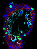
Figure 1. TAMs and OC cells in vitro 3D co-culture system were showed by Immunofluorescence. A. TAMs and OC cells form spheroids in an in vitro 3D co-culture system. GFP+F4/80+CD206+ TAMs isolated from spheroids of ovarian cancer-bearing donor tomatolysM-cre mice and ID8 cells were co-cultured in the Matrigel-precoated 24-well plate for 48 h. The spheroids were subjected to immunofluorescent staining for E-cadherin for tumor cells. Images for GFP+ TAMs, E-Cadherin+ OC cells and DAPI for all cells in the spheroids are shown. B. Human TAMs were isolated and infected with lentivirus expressing RFP. RFP-expressing TAMs were incubated with SKOV3 human ovarian cancer cells followed by 3D co-culture for 72 h. Spheroids were immunostained with keratin-14. Scale bars = 10 μm.
Materials and Reagents
- Pipette tips
- 15 cm Petri dish
- 10 ml serological pipette
- 50 ml sterile conical tube (Corning, Falcon®, catalog number: 352070 )
- 18-gauge needle
- 10 ml syringe
- FalconTM cell strainers,100 μm (Corning, Falcon®, catalog number: 352360 )
- 1.5 ml microcentrifuge tubes
- Greiner cellstar multiwall culture plates (24 wells, Greiner Bio One International, catalog number: 662102 )
- Cell lines: ID8 ovarian cancer cell line (Yin et al., 2016) was a gift from Jack Lawler and Carmelo Nucera at Beth Israel Deaconess Medical Center (Harvard Medical School, Boston, Massachusetts, USA).
Note: ID8 cells are mouse epithelial OC line derived from C57BL/6 background; Passage under 30 and less than 1 week culture before injections. - C57BL/6 mice, female, age: 8 weeks
- Tomato reporter transgenic mice: ID8 OC cells were labeled by stably expressing mCherry fluorescence protein while LysMCre mice crossed to the tomato reporter mT/mG
- Dulbecco’s modified Eagle’s medium (DMEM) (Thermo Fisher Scientific, GibcoTM, catalog number: 10567014 )
- D-glucose
- Fetal bovine serum (FBS) (Thermo Fisher Scientific, GibcoTM, catalog number: 26140079 )
- Penicillin-streptomycin (Thermo Fisher Scientific, GibcoTM, catalog number: 15140 )
- 0.25% Trypsin-EDTA (Thermo Fisher Scientific, GibcoTM, catalog number: 25200056 )
- Ketamine
- Bovine serum albumin (BSA) (Thermo Fisher Scientific, Thermo ScientificTM, catalog number: B14 )
- Collagenase (Thermo Fisher Scientific, GibcoTM, catalog number: 17104019 )
- F4/80 monoclonal antibody, APC conjugate for flow cytometry (Thermo Fisher Scientific, Invitrogen, catalog number: MF48005 )
- PE anti-mouse CD206 (MMR) antibody (BioLegend, catalog number: 141705 )
- DAPI (Vector Laboratories, catalog number: H-1200 )
- Anti-mouse E-Cadherin antibody (BD, PharmingenTM, catalog number: 610404 )
- Donkey anti-mouse (Alexa Flour 594) (Thermo Fisher Scientific, InvitrogenTM, catalog number: A-21203 )
- Live cell tracker CMFDA (Thermo Fisher Scientific, InvitrogenTM, catalog number: C2925 )
- EGFP (abm, catalog number: LV011-a )
- Na2HPO4
- NaCl
- KH2PO4
- KCl
- Corning Matrigel (Basement membrane matrix, Corning, catalog number: 356234 )
- Tween 20 (Sigma-Aldrich, catalog number: P7949-500ML )
- Paraformaldehyde (Sigma-Aldrich, catalog number: 16005 )
- PBS (see Recipes)
- PBST (see Recipes)
- AC buffer (0.5% BSA) (see Recipes)
- 2% Matrigel (see Recipes)
- 3.7% paraformaldehyde (see Recipes)
Equipment
- Safety cabinet
- Pipette-aid
- Centrifuge with swinging-bucket rotor and adaptors for 50-ml conical tubes
- Water bath set at 37 °C
- Humidified cell culture incubator set to 37 °C and 5% CO2
- Zeiss Axiovert 200 fluorescence microscope (Carl Zeiss, model: Axiovert 200 )
- Upright microscope with 10x objective
- Cell sorter and the scale (BD, model: FACSAriaTM II ); sorting at a rate of 80,000 cell/h
Software
- Quantitation of the average distance between the geometrical center of nuclei of adjacent cells can be measured using Openlab3 software (Improvision, Lexington, MA) or other commercially available image analysis software
- SAS software (version 9.1.4, SAS Institute, Cary, NC)
Procedure
文章信息
版权信息
© 2018 The Authors; exclusive licensee Bio-protocol LLC.
如何引用
Long, L., Yin, M. and Min, W. (2018). 3D Co-culture System of Tumor-associated Macrophages and Ovarian Cancer Cells. Bio-protocol 8(8): e2815. DOI: 10.21769/BioProtoc.2815.
分类
免疫学 > 免疫细胞功能 > 巨噬细胞
癌症生物学 > 肿瘤免疫学 > 细胞生物学试验 > 细胞分离和培养
细胞生物学 > 细胞分离和培养 > 3D细胞培养
您对这篇实验方法有问题吗?
在此处发布您的问题,我们将邀请本文作者来回答。同时,我们会将您的问题发布到Bio-protocol Exchange,以便寻求社区成员的帮助。