

Centromere Chromosome Orientation Fluorescent in situ Hybridization (Cen-CO-FISH) Detects Sister Chromatid Exchange at the Centromere in Human Cells
着丝粒染色体定位荧光原位杂交法(Cen-CO-FISH)检测人细胞中着丝粒DNA重复的姐妹染色单体交换事件

发布: 2018年04月05日第8卷第7期 DOI: 10.21769/BioProtoc.2792 浏览次数: 9940
评审: Gal HaimovichShalini Low-NamAnonymous reviewer(s)
参见作者原研究论文
The authors used this protocol in:
Feb 2017
Advertisement


Abstract
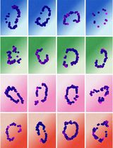
Human centromeres are composed of large tandem arrays of repetitive alpha satellite DNA, which are often sites of aberrant rearrangement in cancers (Mitelman et al., 1997; Padilla-Nash et al., 2001). To date, annotation of the human centromere repetitive sequences remains incomplete, greatly hindering in-depth functional studies of these regions essential for chromosome segregation. In order to monitor sister chromatid exchange happening at the centromere (C-SCE) due to recombination and mutagenic events, I have applied the Chromosome-Orientation Fluorescence in situ Hybridization (CO-FISH) technique to centromeres (Cen-CO-FISH) in human cells. This hybridization-based method involves (1) the incorporation of nucleotide analogs through a single round of replication, (2) enzymatic digestion of the newly synthesized DNA strand and (3) subsequent hybridization of single-stranded probes, in absence of a denaturation step. The resulting signal allows to differentially label each sister chromatid based on the 5’-3’ directionality of the DNA and to score aberrant staining patterns indicative of C-SCE. The Cen-CO-FISH method applied to human centromeres revealed that human centromeres indeed undergo recombination in cycling cells resulting in C-SCE, and centromere instability is enhanced in cancer cell lines and primary cells undergoing senescence (Giunta and Funabiki, 2017). Here, I present the detailed protocol of the preparation, experimental procedure and data acquisition for the Cen-CO-FISH method in human cells. It also includes a conceptual overview of the technique, with examples of representative images and scoring guidelines. The Cen-CO-FISH represents a valuable tool to facilitate exploration of centromere repeats.
Keywords: Centromere (着丝粒)Background
The human genome project was marked completed in 2003, yet it omitted over 10% of the human repetitive DNA (de Koning et al., 2011), including the centromere. The human centromere is a highly specialized genomic locus (Choo, 1997) playing a critical role during chromosome segregation where it serves as the site of kinetochore assembly to allow interaction with microtubules and sister chromatids separation during cell division (Cheeseman, 2014). Human centromeres are made of characteristic repetitive DNA sequences called alpha-satellites, whose linear assembly remains largely absent from the reference genomes. Here, I present the application of the Cen-CO-FISH technique to label human centromere and monitor recombination events resulting in crossover. Introduced by Bailey and colleagues over 20 years ago (Bailey et al., 1996), the CO-FISH method has been widely applied to detect recombination, fragility, replication timing, fusion and inversions at telomeres repeats, as well as to monitor mitotic segregation patterns and non-random sister chromatid segregation (Bailey et al., 2010). The application of this methodology to centromere, hereby called Cen-CO-FISH method, has revealed that the centromere-specific histone variant CENP-A, and CENP-A associated proteins CENP-C and CENP-T/W, work to prevent centromere instability and this functionality is compromised in cancer cell lines and in primary cells approaching replicative senescence that display higher number of C-SCE (Giunta and Funabiki, 2017). Cen-CO-FISH was used to assess centromere instability in cancer and during cellular senescence in human cells (Giunta and Funabiki, 2017) and it has been previously applied to study recombination (Jaco et al., 2008; de La Fuente et al., 2015) and sister chromatid separation patterns in mouse cells (Falconer et al., 2010). The wide application potentials of this methodology spans from quantitative detection of alpha satellite repeats, centromere recombination resulting in C-SCE, fragility, replication timing, fusion and inversions, as well as to monitor mitotic segregation patterns and non-random sister chromatids segregation (Bailey et al., 2010; Yadlapalli and Yamashita, 2013). Cen-CO-FISH fills the gaps in the missing genetic information that have cast a shadow over the centromere and other repetitive regions, bringing new light into the possibilities for functional exploration of these important loci of our genome.
Materials and Reagents
- 6 or 10 cm Petri dish (Corning, Falcon®, catalog numbers: 353002 or 353003 )
- 15 ml Falcon tube (Corning, Falcon®, catalog number: 352097 )
- Frosted slides (Superfrost Plus; Fisher Scientific, catalog number: 12-550-15 )
- Coverslips (24 x 60 mm) (Fisher Scientific, catalog number: 12-545-M )
- Paper towel
- Glass Pasteur pipette (Fisher Scientific, catalog number: 13-678-20A )
- Gloves and lab coat
- Human cells of interest and appropriate medium
Note: This protocol is for adherent cells, changes can be made for use for non-adherent cultures. - 5’-Bromodeoxyuridine (BrdU) (MP Biomedicals, catalog number: 100166 )
Note: Prepare 10 mM stock solution in double distilled water (1,000x); make aliquots and store at -20 °C. - 5’-Bromodeoxycytidine (BrdC) (Sigma-Aldrich, catalog number: B5002 )
Note: Prepare 10 mM stock solution in double distilled water (1,000x); make aliquots and store at -20 °C. - Colcemid (Roche Diagnostics, catalog number: 10295892001 ; already diluted 10 μg/ml)–Store at 4 °C
- Phosphate-buffered saline (PBS)
- Trypsin-EDTA (Thermo Fisher Scientific, GibcoTM, catalog number: 25300 )
- Fetal bovine serum (Atlanta Biologicals)
- Potassium chloride (KCl) (Fisher scientific, catalog number: P217-500 )
- RNase A (Sigma-Aldrich, catalog number: R5000 )
Note: Prepare stock solution 50 mg/ml in 10 mM Tris-HCl pH 7.2. Aliquot and store at -20 °C. - Hoechst 33258 (Thermo Fisher Scientific, InvitrogenTM, catalog number: H3569 )
Note: Make a 10 μg/ml solution in double distilled water and store at 4 °C away from light. - Exonuclease III and buffer (Promega, catalog number: M1811 )–Keep at -20 °C
- DAPI (Sigma-Aldrich, catalog number: D9542 )–0.5 mg/ml stock in water. Keep at 4 °C in the dark for one year
- ProLong Gold Anti-fade Reagent (Thermo Fisher Scientific, InvitrogenTM, catalog number: P36934 )
- Nail varnish (Sally Hansen, Transparent Harderer)
- Methanol (Fisher Scientific, catalog number: A452-4 )
- Glacial acetic acid (Fisher Scientific, catalog number: A38C-212 )
- Ethanol 100% (Decon, catalog number: 2716 ), 90% and 70%
- Blocking reagent (Roche Diagnostics, catalog number: 11096176001 )
- Maleic acid (Sigma-Aldrich, catalog number: M0375 )
- Sodium chloride (NaCl) (Merck, catalog number: SX0420-5 )
- Sodium hydroxide (NaOH) (Fisher Scientific, catalog number: S318-500 )
- Tris-HCl pH 7.2 (Sigma-Aldrich, CAS number: 1185-53-1)
- Formamide (Fisher Scientific, catalog number: BP228 ; use deionized for hybridization)
- Bovine serum albumin (BSA)
- Tween-20 (Hoefer, CAS number: GR128-500)
- Sodium citrate (Sigma-Aldrich, CAS number: 6132-04-3)
- Magnesium chloride (MgCl2) (Fisher Scientific, catalog number: AC41341-5000)
Manufacturer: Acros Organics, catalog number: 413410025 . - Dithiothreitol (DTT) (Thermo Fisher Scientific, Thermo ScientificTM, catalog number: R0861 )
- Hypotonic solution (see Recipes)
- Fixative solution (see Recipes)
- Ethanol dilution (see Recipes)
- Blocking solution (see Recipes)
- Hybridization solution (see Recipes)
- Hybridization wash #1 (see Recipes)
- Hybridization wash #2 (see Recipes)
- Peptide nucleic acid (PNA) probes (custom probes from PNABio) (see Recipes)
- Sodium chloride and sodium citrate buffer (SSC, see Recipes)
Equipment
- Pipettes (Gilson)
- Centrifuge (Eppendorf, model: 5810 R )
- Coplin Jars (Scienceware, Sigma-Aldrich, catalog number: S5641 )
- Heating block (VWR)
- Water bath (Fisher Scientific, model: IsotempTM 210 )
- Stratalinker with 365-nm UV light blubs (Spectralinker XL-1000 1800 UV irradiator) (Spectronics Corporation, model: XL-1000 )
- Slides plastic tray–to fit into the Stratalinker
- Hybridization chamber (see text for more details)
- Orbital shaker
- Imaging equipment:
- DeltaVision Image Restoration microscope system (Applied Precision/GE Healthcare)
- Olympus IX-70 microscope (Olympus, model: IX70 )
- 100x/1.40 UPLSAPO objective lens
- CoolSnap QE CCD camera (Photometrics)
- DeltaVision Image Restoration microscope system (Applied Precision/GE Healthcare)
Software
- SoftWoRx (Sold by Applied Precision)
- Metamorph 7.8 (Sold by Universal Imaging)
- Prism 5 (Sold by GraphPad)
Procedure
文章信息
版权信息
© 2018 The Authors; exclusive licensee Bio-protocol LLC.
如何引用
Giunta, S. (2018). Centromere Chromosome Orientation Fluorescent in situ Hybridization (Cen-CO-FISH) Detects Sister Chromatid Exchange at the Centromere in Human Cells. Bio-protocol 8(7): e2792. DOI: 10.21769/BioProtoc.2792.
分类
癌症生物学 > 基因组不稳定性及突变 > 遗传学
癌症生物学 > 通用技术 > 免疫学试验
细胞生物学 > 细胞成像 > 荧光
您对这篇实验方法有问题吗?
在此处发布您的问题,我们将邀请本文作者来回答。同时,我们会将您的问题发布到Bio-protocol Exchange,以便寻求社区成员的帮助。