

Measurement of Intracellular ROS in Caenorhabditis elegans Using 2’,7’-Dichlorodihydrofluorescein Diacetate
使用2',7'-二氯二氢荧光素二乙酸盐测定秀丽隐杆线虫细胞内ROS
.jpg)
发布: 2018年03月20日第8卷第6期 DOI: 10.21769/BioProtoc.2774 浏览次数: 16737
评审: Neelanjan BoseMichael EnosAnonymous reviewer(s)
参见作者原研究论文
The authors used this protocol in:

Oct 2017
Advertisement


Abstract
Reactive oxygen species (ROS) are generated during normal metabolic processes under aerobic conditions. Since ROS production initiates harmful radical chain reactions on cellular macromolecules, including lipid peroxidation, DNA mutation, and protein denaturation, it has been implicated in a wide spectrum of diseases such as cancer, cardiovascular disease, ischemia-reperfusion and aging. Over the past several decades, antioxidants have received explosive attention regarding their protective potential against these deleterious reactions. Accordingly, many analytical methodologies have been developed for the evaluation of the antioxidant capacity of compounds or complex biological samples. Herein, we introduce a simple and convenient method to detect in vivo intracellular ROS levels photometrically in Caenorhabditis elegans using 2’,7’-dichlorofluorescein diacetate (H2DCFDA), a cell permeant tracer.
Keywords: Reactive oxygen species (活性氧族)Background
In situ detection of intracellular reactive oxygen species (ROS) levels in the living organism using fluorescent probe 2’,7’-dichlorofluorescein diacetate (H2DCFDA) has been broadly performed by those researchers who work in the field of oxidative stress and related diseases. The non-polar and non-ionic probe, H2DCFDA, can easily penetrate the cellular membrane and is enzymatically deacetylated by esterases. This biochemical reaction turns H2DCFDA into the non-fluorescent compound H2DCF which is then rapidly oxidized to highly fluorescent 2’,7’-dichlorofluorescein (DCF) in the presence of ROS (Figure 1A). Therefore, fluorescence signals from H2DCFDA probe demonstrate important information for the quantification of ROS at single cell level (Labuschagne and Brenkman, 2013). The Caenorhabditis elegans model system provides an excellent in vivo experimental environment for evaluating molecular mechanisms of ROS pathophysiology due to their short lifespan, simplicity, and ease of genetic manipulation (Labuschagne and Brenkman, 2013; Miranda-Vizuete and Veal, 2017; Yoon et al., 2017) (Figure 1B). Here, we describe a simple protocol to measure the levels of time-course ROS generation in C. elegans using H2DCFDA under normal and heat- or chemically-induced oxidative stress conditions. Using this protocol, we determined the effects of H2DCFDA concentration and number of tested worms on DCF fluorescence signal (Figure 2).
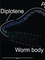
Figure 1. ROS detection. A. Production of florescent DCF by intracellular ROS. B. Measurement of intracellular ROS using molecular probe (H2DCFDA) in C. elegans.
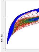
Figure 2. Change in DCF fluorescence signals depends on experimental conditions. A. The expression of gcs-1(promoter)::GFP (oxidative stress marker) transgene by oxidative stress. Heat (30 °C for 2 h) induced the expression of gcs-1(promoter)::GFP transgene in the intestines of live nematodes. B. Concentration-response curves were plotted in terms of the mean value of DCF fluorescence signals induced by 12.5, 25, 50, and 100 μM of H2DCFDA using 50 nematodes for each experiment. C. Changes in DCF fluorescence signals depending on the number of tested nematodes (10, 20, 50, and 100) were assessed using 25 μM of H2DCFDA.
Materials and Reagents
- 1.5 ml microcentrifuge tube (Corning, Axygen®, catalog number: MCT-150-C )
- Conical tube, 15 ml (Corning, Falcon®, catalog number: 352096 )
- Slide glass (VWR, catalog number: 48300-025 )
- 96-well microplate, black (SPL Life Sciences, catalog number: 30496 )
- 60 mm Petri dish (SPL Life Sciences, catalog number: 10060 )
- Platinum wire, 0.2 mm (Alfa Aesar, catalog number: 45093 )
- Caenorhabditis elegans strain, wild-type [N2] (Caenorhabditis Genetics Center, University of Minnesota: https://cgc.umn.edu)
- Escherichia coli OP50 strain (Caenorhabditis Genetic Center)
- Distilled water
- 2’,7’-Dichlorofluorescein diacetate (Sigma-Aldrich, catalog number: D6883 )
- Dimethyl sulfoxide (DMSO) (Sigma-Aldrich, catalog number: D2650 )
- Methyl viologen dichloride hydrate (Merck, catalog number: 856177 )
- Cholesterol (Sigma-Aldrich, catalog number: C8667 )
- Ethanol (Sigma-Aldrich, catalog number: E7023 )
- Sodium chloride (NaCl) (Sigma-Aldrich, catalog number: S5886 )
- Bacto-peptone (BD, BactoTM, catalog number: 211677 )
- Agar (Sigma-Aldrich, catalog number: A1296 )
- Calcium chloride (CaCl2) (Sigma-Aldrich, catalog number: C1016 )
- Magnesium sulfate (MgSO4) (Sigma-Aldrich, catalog number: M7506 )
- Potassium phosphate, dibasic (K2HPO4) (Sigma-Aldrich, catalog number: P3786 )
- Potassium phosphate, monobasic (KH2PO4) (Sigma-Aldrich, catalog number: 795488 )
- Sodium phosphate dibasic (Na2HPO4) (Sigma-Aldrich, catalog number: S3264 )
- Sodium hydroxide (NaOH) (Sigma-Aldrich, catalog number: S8045 )
- Sodium hypochlorite (NaClO) (Sigma-Aldrich, catalog number: 425044 )
- 5 mg/ml cholesterol (see Recipes)
- Nematode growth medium (NGM) agar plates (see Recipes)
- M9 buffer (see Recipes)
- Bleaching solution (see Recipes)
Equipment
- Shaking incubator (Benchtop Shaking Incubator, VWR, model: Model 1570 )
- Micropipette (VWR International)
- Incubators for stable temperature (VWR SIGNATURE 20-cuft Model 2020 B.O.D. -10 °C to 45 °C Low Temp Incubator) (VWR, Advanced Instruments, model: Model 2020 )
- Tabletop centrifuge (VWR, model: VWR® Mini Centrifuge C1413V )
- Dissecting stereomicroscope (Stereo Microscope with Apochromatic Optics Leica S8 APO) (Leica Microsystems, model: Leica S8 APO )
- Fluorophotometer (Promega, model: GloMax®-Multi Microplate Multimode Reader )
- Vortex mixer (Standard Heavy-Duty Vortex Mixer, VWR, catalog number: 97043-562 )
- Autoclave (Hanshin Medical, model: HS-2321SD )
Software
- Microsoft Office 2017 Excel (Microsoft Corporation, Redmond, USA)
- IBM SPSS statistics 24 (IBM Corporation, New York, USA)
Procedure
文章信息
版权信息
© 2018 The Authors; exclusive licensee Bio-protocol LLC.
如何引用
Yoon, D. S., Lee, M. and Cha, D. S. (2018). Measurement of Intracellular ROS in Caenorhabditis elegans Using 2’,7’-Dichlorodihydrofluorescein Diacetate. Bio-protocol 8(6): e2774. DOI: 10.21769/BioProtoc.2774.
分类
发育生物学 > 细胞信号传导 > 胁迫反应
生物化学 > 其它化合物 > 氧
细胞生物学 > 细胞信号传导 > 胞内信号传导
您对这篇实验方法有问题吗?
在此处发布您的问题,我们将邀请本文作者来回答。同时,我们会将您的问题发布到Bio-protocol Exchange,以便寻求社区成员的帮助。