

Determination of DNA Damage in the Retina Photoreceptors of Drosophila
果蝇视网膜光感受器DNA损伤测定
.JPG)
发布: 2018年02月05日第8卷第3期 DOI: 10.21769/BioProtoc.2708 浏览次数: 7136
评审: Oneil G. BhalalaSandhya GanesanAnonymous reviewer(s)
参见作者原研究论文
The authors used this protocol in:
Sep 2017
Advertisement


Abstract
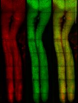
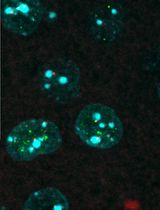
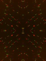
The retina is sensitive for light damages, because of direct light exposure, especially intense blue and UV light, which increase level of ROS and other toxic phototransduction products in photoreceptor cells. In our previous work (Damulewicz et al., 2017a and 2017b), we used 8-oxo-deoxyguanosine (8-OHdG) as a marker for oxidative stress to investigate the role of heme oxygenase in DNA protection against UV light. In this protocol, we showed how to determine the level of DNA damages in the retina using immunohistochemical staining.
Keywords: Oxidative stress (氧化应激)Background
Deoxyribonucleic acid (DNA) is an essential molecule for all living organisms. It contains genetic code with instruction for proteins and other molecules necessary for structure and metabolism of an organism. DNA is composed of two strands of nucleotides. Each nucleotide is built of one of four types of nucleobases (cytosine, guanine, adenine and thymine), deoxyribose and phosphate group. Physical and chemical changes in DNA structure are classified as DNA damages. It includes one or two strands of DNA breaks, missing nucleotide or chemical changes of nucleobases. Damages can be induced by environmental factors, like chemicals or UV light exposure, or in result of metabolic processes, which produce reactive oxygen species, reactive nitrogen species, reactive carbonyl species and alkylating agents (De Bont and van Larebeke, 2004).
One of the most common DNA damage is 8-oxo-deoxyguanosine (8-OHdG), which is a product of DNA oxidation by reactive oxygen species and it serves as a marker of oxidative stress (De Souza-Pinto et al., 2001; Swenberg et al., 2011; Valavanidis et al., 2013). 8-OHdG enhances the risk of transversion mutation, because it has ability to pair with adenine and cytosine bases (Maki and Sekiguchi, 1992). In effect it plays a role in mutagenesis, cancerogenesis and aging (De Bont and van Larebeke, 2004; Kohen and Nyska, 2002). Higher 8-OHdG level was described in breast, renal and gastric tumors (Lee et al., 1998; Musarrat et al., 1996; Okamoto et al., 1994).
There are several methods to determine 8-OHdG level in a specific tissue, like immunohistochemistry (De Carvalho et al., 2013), Western blot (Kannan et al., 2006), standard HPLC (Herrero and Barja, 1999), liquid chromatography nanoelectrospray-tandem mass spectrometry (Ma et al., 2016). Here, we present a detailed protocol for DNA damage determination in the retina of fruit fly, Drosophila melanogaster using immunohistochemistry method (Damulewicz et al., 2017a and 2017b).
Materials and Reagents
- Filter paper (Fisher Scientific, catalog number: 09-790-14D )
- Cover glasses (Menzel-Glaser)
- Microscope glasses (Superfrost, Menzel-Glaser)
- Small plastic holders (i.e., Eppendorf tube lids)
- Tissue-Tek (SAKURA, catalog number: 4583 )
- Drosophila melanogaster (wild-type CantonS, 5-7 days old males)
Note: Local ethical standards need to be followed.
- 6% glucose in water (commercial)
- Protoporphyrin IX (SnPP IX) (Frontier Scientific, catalog number: P562-9 )
- Hemin chloride (Merck, Calbiochem, catalog number: 3741 )
- CO2
- Schneider’s medium (Sigma-Aldrich, catalog number: S9895 )
- Etoposide (Sigma-Aldrich, catalog number: E1383 )
- 4% paraformaldehyde (PFA) (Sigma-Aldrich, catalog number: P6148 )
- Sucrose (commercial)
- Liquid nitrogen
- Normal Goat Serum (NGS) (Sigma-Aldrich, catalog number: G9023 )
- Bovine serum albumin (BSA)
- Mouse anti-8-hydroxyguanosine primary antibodies (Acris, catalog number: AM03160 )
- HRP/DAB (ABC) kit (Abcam, catalog number: ab64264 )
- Glycerol (MP Biomedicals)
- Vectashield with DAPI (Vector Laboratories, catalog number: H-1200 )
- Sodium chloride (NaCl) (Sigma-Aldrich, catalog number: S7653 )
- Potassium chloride (KCl) (Sigma-Aldrich, catalog number: P9541 )
- Sodium phosphate monobasic monohydrate (NaH2PO4·H2O) (Sigma-Aldrich, catalog number: S3522 )
- Potassium phosphate monobasic (KH2PO4) (Sigma-Aldrich, catalog number: P5655 )
- Hydrochloric acid (HCl) (Sigma-Aldrich, catalog number: H1758 )
- Sodium phosphate dibasic (Na2HPO4) (Sigma-Aldrich, catalog number: S7907 )
- Triton X-100 (Sigma-Aldrich, catalog number: T8787 )
- Cornmeal (Glutenex)
- Agar (Bioshop)
- Artificial honey (Vortumnus)
- Yeast (Paneo)
- Methyl-4-hydroxybenzoate (anti-fungal chemical, Sigma-Aldrich, catalog number: H3647 )
- 90% EtOH
- Phosphate buffered saline (PBS) (see Recipes)
- Phosphate buffer with Triton (PBT) (see Recipes)
- Standard medium for flies (see Recipes)
Equipment
- Forceps (Dumont No. 5)
- UV transilluminator (Vilbert Lourmat, model: UVECX26 )
- White light transilluminator (Vilbert Lourmat, model: TFX53WL )
- Cryostat (Leica, model: CM1850 UV )
- Light microscope (Carl Zeiss, model: Axioskop 50 )
- Camera (Canon, model: G10 )
Software
- ImageJ software
Procedure
文章信息
版权信息
© 2018 The Authors; exclusive licensee Bio-protocol LLC.
如何引用
Damulewicz, M. and Pyza, E. (2018). Determination of DNA Damage in the Retina Photoreceptors of Drosophila. Bio-protocol 8(3): e2708. DOI: 10.21769/BioProtoc.2708.
分类
分子生物学 > DNA > DNA 损伤和修复
神经科学 > 神经解剖学和神经环路 > 视神经
您对这篇实验方法有问题吗?
在此处发布您的问题,我们将邀请本文作者来回答。同时,我们会将您的问题发布到Bio-protocol Exchange,以便寻求社区成员的帮助。