

Micro-computed Tomography to Visualize Vascular Networks in Maize Stems
玉米茎杆中维管网络的微型计算机断层摄影术观察


发布: 2018年01月05日第8卷第1期 DOI: 10.21769/BioProtoc.2682 浏览次数: 10409
评审: Annis Elizabeth RichardsonRenate WeizbauerAnonymous reviewer(s)
参见作者原研究论文
The authors used this protocol in:
Jun 2017


Abstract
Plant vascular systems in the stem connect roots with aerial organs to move solutes containing minerals, nutrients as well as signaling molecules, and therefore, they play pivotal roles in plant growth and development. However, stem vascular systems, especially in crop species, have been poorly described since they are deeply embedded in the tissue. Here we describe a protocol to utilize micro-computed tomography (micro-CT) scanning to visualize vascular networks in the maize stem. The protocol covers sample fixation and staining with contrasting reagents, data acquisition using micro-CT, reconstructing three-dimensional (3D) models of stem inner structures and extraction of vascular networks from the model. This protocol can be easily applied to various types of species and organs/tissues.
Keywords: Micro-CT scanning (微型CT扫描)Background
Monocot stems have a characteristic vascular network in which veins remain separate and independent. Despite its importance to supporting growth and development, the pattern of vascular networks in monocot stems has been poorly studied. Visualization of stem vascular networks is quite challenging because veins are deeply embedded in tissues. Conventional tissue sectioning can be applied to observe the networks, however, it is a laborious and time-consuming process which requires observation of hundreds of sections. In addition, the size of crop stems, which is much larger than the field of view under the microscopes, makes it difficult to capture the whole system.
Recently, we reported that maize transcription factor BEL1-like homeobox (BLH) 12 and BLH14 play important roles in the stem development and vein network formation (Tsuda et al., 2017). To obtain a comprehensive view of the vascular systems, we adopted the micro-CT scanning described previously and optimized it for maize stems (Metscher, 2009a and 2009b, Degenhardt et al., 2010, Staedler et al., 2013, Gignac et al., 2016). By combining this method with image analyses, we were able to reconstruct 3D models of inner stem structures in an efficient and reliable manner. This protocol can be used to visualize inner structures such as veins in various species and tissues.
Materials and Reagents
- 50 ml Polypropylene tube (Greiner Bio One International, catalog number: 227261 )
- Kimwipes
- 1.5 ml micro-tube (Eppendorf, catalog number: 0030125150 )
- Filter foams MOLTOFILTER MF-13 thickness 5 mm (INOAC Corp.)
- Maize (B73)
- Formalin (Wako Pure Chemical Industries, catalog number: 061-00416 )
- Acetic acid (Wako Pure Chemical Industries, catalog number: 017-00256 )
- Ethanol (Wako Pure Chemical Industries, catalog number: 057-00451 )
- Lugol stock solution
- Potassium iodide (Wako Pure Chemical Industries, catalog number: 166-03971 )
- Iodine (Wako Pure Chemical Industries, catalog number: 094-05421 )
- Fixative FAA solution (see Recipes)
- Iodine staining (Degenhardt et al., 2010) (see Recipes)
- 25% Lugol working solution (see Recipes)
Equipment
- X-ray micro-CT imaging system (Comscantechno, model: ScanXmate-E090S105 ) (Figure 1)
- X-ray tube: Microfocus X-ray source (Hamamatsu Photonics K.K., model: L9421-02 )
- Detector: Flat panel detector (Varex Imaging, model: PaxScan 1313DX )
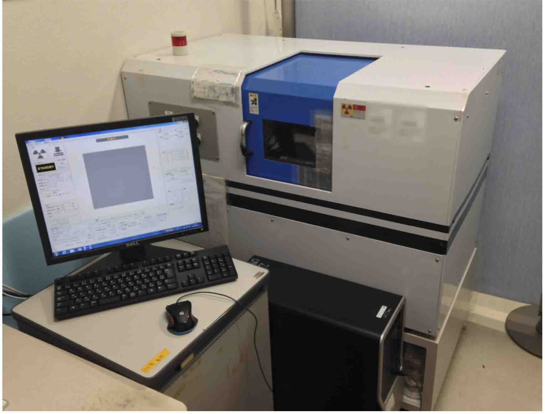
Figure 1. X-ray micro-CT imaging system used in this protocol
- X-ray tube: Microfocus X-ray source (Hamamatsu Photonics K.K., model: L9421-02 )
Software
- coneCTexpress (built in the micro-CT imaging system. Comscantechno Co. Ltd., Kanagawa, Japan)
- OsiriX MD v8.5 (http://www.osirix-viewer.com, Pixmeo SARL, Swiss)
- Imaris 8.2 (http://www.bitplane.com, Bitplane, UK)
- Adobe Premiere Pro CC (http://www.adobe.com, Adobe, US)
Procedure
文章信息
版权信息
© 2018 The Authors; exclusive licensee Bio-protocol LLC.
如何引用
Maeno, A. and Tsuda, K. (2018). Micro-computed Tomography to Visualize Vascular Networks in Maize Stems. Bio-protocol 8(1): e2682. DOI: 10.21769/BioProtoc.2682.
分类
植物科学 > 植物发育生物学 > 综合
细胞生物学 > 组织分析 > 组织成像
您对这篇实验方法有问题吗?
在此处发布您的问题,我们将邀请本文作者来回答。同时,我们会将您的问题发布到Bio-protocol Exchange,以便寻求社区成员的帮助。