

Ex vivo Trophoblast-specific Genetic Manipulation Using Lentiviral Delivery
使用慢病毒递送的离体滋养层特定基因操作

发布: 2017年12月20日第7卷第24期 DOI: 10.21769/BioProtoc.2652 浏览次数: 8161
评审: Giusy TornilloIrit AdiniAnonymous reviewer(s)
参见作者原研究论文
The authors used this protocol in:
Nov 2016
Advertisement


Abstract
In this protocol report, we describe a lentiviral gene delivery technique for genetic modification of the rat trophoblast cell lineage. Lentiviral packaged gene constructs can be efficiently and specifically delivered to the trophoblast cell lineage of the blastocyst. The consequences of ‘gain-of-function’ and ‘loss-of-function’ blastocyst manipulations can be evaluated with in vitro outgrowth assays or following transfer to pseudopregnant rats.
Keywords: Blastocyst (囊胚)Background
The placenta functions as a conduit between the mother and the developing fetus and is essential for viviparity (Georgiades et al., 2002). It is specialized to facilitate and coordinate maternal adaptations to pregnancy and fetal development (Soares et al., 2014; Burton et al., 2016). The placenta contains trophoblast cells, which perform several specialized functions. Acquisition of trophoblast cell specializations requires highly regulated differentiation of trophoblast stem and progenitor cell populations (Maltepe and Fisher, 2015). The mature rat placenta is comprised of two morphologically and functionally distinct compartments: the junctional zone and the labyrinth zone (Soares et al., 2012). Progenitor cells, spongiotrophoblast cells, glycogen trophoblast cells, and polyploid trophoblast giant cells comprise the junctional zone. An invasive trophoblast lineage arises from progenitors within the junctional zone region (Ain et al., 2003; Soares et al., 2014). During the last week of gestation, these cells move out of the placenta and invade into the maternal uterine mesometrial compartment (Ain et al., 2003; Pijnenborg and Vercruysse, 2010). The innermost layer of the placenta (proximal to the developing fetus) is called the labyrinth zone, which consists of trophoblast cells and fetal vasculature derived from the allantois. Progenitor trophoblast cells within the labyrinth zone fuse to form syncytia, which provide barriers between maternal and fetal compartments (Soares et al., 2012). The labyrinth zone consists of an elaborate branched structure, providing a large surface area for nutrient, waste and gas exchange with the fetus (Knipp et al., 1999; Watson and Cross, 2005).
Specific modifications of the trophoblast lineage can be achieved using lentiviral transduction of the outer layer of the embryo at the blastocyst stage, termed trophectoderm (Georgiades et al., 2007; Malashicheva et al., 2007; Okada et al., 2007; Lee et al., 2009) (Figure 1). This method allows for efficient manipulation of all trophoblast cell lineages. It also facilitates discrimination between trophoblast and embryonic contributions to phenotypic outcomes arising from global genetic manipulations. The experimental basis for selective trophoblast viral infection is directly related to the structure of the blastocyst and the tight epithelium formed by the outer trophectoderm, which effectively restricts viral particle access to the inner cell mass (Georgiades et al., 2007; Malashicheva et al., 2007; Okada et al., 2007; Lee et al., 2009). This method can be utilized to investigate the consequences of both ‘gain-of-function’ and ‘loss-of-function’ manipulations of the trophoblast lineage, which can provide insights into molecular mechanisms controlling placental development (Georgiades et al., 2007; Malashicheva et al., 2007; Okada et al., 2007; Lee et al., 2009; Morioka et al., 2009; Kent et al., 2011; Chakraborty et al., 2016; Muto et al., 2016).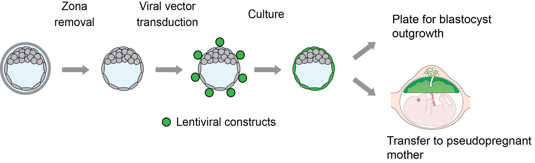
Figure 1. Schematic showing experimental plan for lentiviral transduction of blastocysts and downstream analyses. Blastocysts are transduced with lentiviral particles (shown in green) expressing specific ‘gain-of-function’ or ‘loss-of-function’ constructs and cultured for 72 h for outgrowth assays or directly transferred to day 3.5 pseudopregnant animals for in vivo analyses.
Materials and Reagents
- Multi-well glass agglutination plates (Scientific Device Laboratory, catalog number: 074-3032B )
- Tissue culture plate, 6-well (MIDSCI, catalog number: TP92006 )
- Needle,18 gauge (BD, BD Biosciences, catalog number: 305195 )
- Syringe, 1 ml
- Tissue culture plate, 12-well (MIDSCI, catalog number: TP92012 )
- Glass mouth pipette (Drummond Scientific, catalog number: 2-000-100 )
- Aspirator tube assembly, 15” (Drummond Scientific, catalog number: 2-000-000 )
- Tissue culture plate, 4-well (Thermo Fisher Scientific, Thermo ScientificTM, catalog number: 176740 )
- Plastic non-tissue culture Petri plates (Fisher Scientific, catalog number: AS4052 )
- Female rats (8-10 weeks of age)
- Lentiviral packaging reagents: pRSV-Rev (Addgene, catalog number: 12253 ), pMDLg/pRRE (Addgene, catalog number: 12251 ), VSVG envelope plasmid (pMD2.G, Addgene, catalog number: 12259 )
- Sterile saline solution (0.9% Sodium Chloride)
- Opti-MEM culture medium (Thermo Fisher Scientific, catalog number: 51985034 )
- Lipofectamine 2000 (Thermo Fisher Scientific, InvitrogenTM, catalog number: 11668-027 )
- p24 enzyme-linked immunoassay kit (Takara Bio, Clontech, catalog number: 632200 )
- M2 medium (Millipore Sigma, catalog number: MR-015-D )
- KSOM medium (Millipore Sigma, catalog number: MR-121-D )
- Acid Tyrode’s solution (Sigma-Aldrich, catalog number: T1788 )
- Mineral oil (Sigma-Aldrich, catalog number: M8410 )
- 4% paraformaldehyde (Sigma-Aldrich, catalog number: 158127 )
- RPMI 1640 (Thermo Fisher Scientific, GibcoTM, catalog number: 11875093 )
- Fetal bovine serum, heat inactivated (Sigma-Aldrich, catalog number: F2442 )
- 2-Mercaptoethanol (Sigma-Aldrich, catalog number: M3148 )
- Sodium pyruvate (Thermo Fisher Scientific, GibcoTM, catalog number: 11360070 )
- Penicillin, and streptomycin (Thermo Fisher Scientific, GibcoTM, catalog number: 15140122 )
- (Optional) Clontech LentiX titration kit (Takara Bio, Clontech, catalog number: 631235 )
- Outgrowth culture medium (see Recipes)
Equipment
- Centrifuge (Beckman Coulter, model: OptimaTM L-100XP , catalog number: 392050)
- Standard inverted light microscope or stereo microscope
- Standard CO2 cell culture incubator, 5% CO2, 37 °C
Software
- ImageJ software (NIH)
Procedure
文章信息
版权信息
© 2017 The Authors; exclusive licensee Bio-protocol LLC.
如何引用
Chakraborty, D., Muto, M. and Soares, M. J. (2017). Ex vivo Trophoblast-specific Genetic Manipulation Using Lentiviral Delivery. Bio-protocol 7(24): e2652. DOI: 10.21769/BioProtoc.2652.
分类
发育生物学 > 细胞生长和命运决定 > 囊胚
细胞生物学 > 细胞工程 > 慢病毒递送
细胞生物学 > 组织分析 > 组织分离
您对这篇实验方法有问题吗?
在此处发布您的问题,我们将邀请本文作者来回答。同时,我们会将您的问题发布到Bio-protocol Exchange,以便寻求社区成员的帮助。