

Proximal Ligation Assay (PLA) on Lung Tissue and Cultured Macrophages to Demonstrate Protein-protein Interaction
对肺组织和培养的巨噬细胞进行邻位连接分析(PLA)以验证蛋白质 - 蛋白质相互作用
发布: 2017年11月05日第7卷第21期 DOI: 10.21769/BioProtoc.2602 浏览次数: 11995
评审: Ivan ZanoniAnonymous reviewer(s)
参见作者原研究论文
The authors used this protocol in:

Jun 2015
Advertisement


Abstract
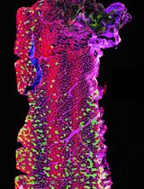
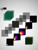
In this protocol, we describe proximal ligation assay (PLA), an antibody-based detection method for protein-protein interaction. This method relies on specific binding of individual primary antibodies to the two putative interacting proteins. The primary antibodies need to have different hosts. The secondary antibodies against the two hosts have complementary oligonucleotide moieties attached to them. If the two antigens are in close proximity (presumably interacting with each other), the complementary oligonucleotides can anneal and fluorescent nucleotides can be incorporated in a single DNA polymerization step. Under a microscope, these reactions appear as punctate fluorescent spots, indicating successful PLA reaction and suggesting protein-protein interaction between the two antigens.
Keywords: Proximal ligation assay (邻位连接分析)Background
Proximal ligation assay (PLA) is an antibody-based technique to determine whether two proteins are with 40 nm of each other. Proteins detected in this manner are identifiable by fluorescence (Ho et al., 2012; Banerjee et al., 2015). This makes PLA an excellent tool for locating protein-protein interactions. Activation of toll-like receptor (TLR) pathways is an important part of the innate immune response to pathogenic threats. TLRs recognize pathogen-associated molecules and induce a signaling cascade to effect a rapid response to infection. TLR2 and TLR4 are two well-studied members of the TLR family that respond to different stimuli. While both receptors activate in response to bacterial infection, only TLR4 responds to lipopolysaccharide exposure. They activate some shared signaling cascades, however, including the MyD88/Traf6 pathway. The induction of this pathway includes the formation a signaling complex known as the myddosome (Gay et al., 2011; Xiong et al., 2011; Cleaver et al., 2014), a protein complex that includes the MyD88, IRAK1, IRAK4 and Traf6 among others. Myddosome assembly results in NF-κb-mediated inflammatory response and pathogen clearance.
Visualizing the engagement of TLR signaling pathways is an important step in identifying and locating immune response. Here, we use PLA to detect TLR pathway activation in fixed lung tissue and a cultured peritoneal macrophage cell-line under treatment with LPS or exposure to opportunistic infection. This method fluorescently labels proteins that interact and remain within close proximity. Using fluorescence microscopy to visualize the resulting labels in vivo allows us to identify the protein complex in respect to tissue location. Here, we demonstrate the ability of this assay to detect TLR2 activation during opportunistic lung infection in vivo and myddosome formation after LPS treatment of peritoneal macrophage cells in vitro. We have also shown the specificity of the technique, as it does not indicate TLR2 activation after LPS treatment in vivo.
Materials and Reagents
- NuncTM Lab-TekTM II Chamber SlideTM System (Thermo Fisher Scientific, Thermo ScientificTM, catalog number: 154534 )
- 0.22 µm PVDF syringe filter (EMD Millipore, catalog number: SLGV004SL )
- Coverslip (22 x 40 mm) (VWR, catalog number: 470019-008 )
- Lung tissue harvested from morphine-treated and/or Streptococcus pneumoniae-infected mice
- J774 cells, a murine peritoneal macrophage cell line (ATCC, catalog number: TIB-67 )
- Primary antibodies
Note: Each pair used in PLA reaction needs to be validated for specificity with Western blot (both native and SDS-PAGE) and must come from a different host, compatible with Duolink secondary antibodies with PLA probes.- Affinity isolated anti-TRAF6 antibody raised in rabbit (Sigma-Aldrich, catalog number: SAB2102531 )
Note: This product has been discontinued.
- Affinity isolated anti-TLR2 antibody raised in rabbit (Sigma-Aldrich, catalog number: SAB1300199 )
- Monoclonal anti-MYD88 (Clone OTI2B2) (OriGene Technologies, catalog number: TA502117 )
- Affinity isolated anti-TRAF6 antibody raised in rabbit (Sigma-Aldrich, catalog number: SAB2102531 )
- Secondary antibodies linked to PLA probes
- Formalin solution, neutral buffered, 10% (10% NBF) (Sigma-Aldrich, catalog number: HT501128-4L )
- Paraffin (Fisher Scientific, catalog number: P31-500 )
- Xylene (Histological grade) (Fisher Scientific, catalog number: X3S-4 )
- Ethanol 200 proof (Merck, catalog number: AX0441 )
- Fixation-permeabilization buffer set (Thermo Fisher Scientific, eBiosciencesTM, catalog number: 88-8824-00 )
- Phosphate-buffered saline (PBS) pH 7.4 (1x) (Thermo Fisher Scientific, GibcoTM, catalog number: 10010023 )
- Tween® 20 (Fisher Scientific, catalog number: BP337-100 )
- Duolink® in situ mounting medium with DAPI (Sigma-Aldrich, catalog number: DUO82049 )
- Nail polish (as cover slip sealant)
- DMEM/High glucose (4,500 mg/L L-glucose) (GE Healthcare, HyCloneTM, catalog number: SH30243.01 )
- Penicillin-streptomycin (10,000 U/ml) (Thermo Fisher Scientific, GibcoTM, catalog number: 15140122 )
- Fetal bovine serum (FBS), qualified, USDA-approved regions (Thermo Fisher Scientific, GibcoTM, catalog number: 10437010 )
- Ultrapure 0.5 M EDTA, pH 6.0 (Thermo Fisher Scientific, InvitrogenTM, catalog number: 15575020 )
- Lipopolysaccharides from Escherichia coli O127:B8 (Sigma-Aldrich, catalog number: L3880 )
- Duolink® in situ wash buffers, fluorescence (Sigma-Aldrich, catalog number: DUO82049 )
- Duolink® in situ detection reagents orange (Sigma-Aldrich, catalog number: DUO92007 )
- Duolink in situ wash buffers A and B (Sigma-Aldrich, catalog number: DUO82047 )
- Duolink in situ wash buffer A working solution (see Recipes)
- Duolink in situ wash buffer B working solution (see Recipes)
Equipment
- Coplin Jar (Generic)
- Oil marker (Aqua-hold pap pen) (Electron Microscopy Sciences, catalog number: 71311 )
- Vegetable steamer (Generic)
- Slide humidity chamber (Simport, model: M920 )
- Laboratory shaker or rocker
- Flourescence filters (Leica)
- Filter set 49, Excitation G365, Emission 445/50
- Filter set 43 HE, Excitation BP550/25, Emission 605/70
- Filter set 49, Excitation G365, Emission 445/50
- Flourescence microscope
Software
- ImageJ software (https://imagej.nih.gov/ij/)
Procedure
文章信息
版权信息
© 2017 The Authors; exclusive licensee Bio-protocol LLC.
如何引用
Mendez, R. and Banerjee, S. (2017). Proximal Ligation Assay (PLA) on Lung Tissue and Cultured Macrophages to Demonstrate Protein-protein Interaction. Bio-protocol 7(21): e2602. DOI: 10.21769/BioProtoc.2602.
分类
免疫学 > 免疫细胞成像 > 共聚焦显微镜技术
细胞生物学 > 细胞成像 > 共聚焦显微镜
您对这篇实验方法有问题吗?
在此处发布您的问题,我们将邀请本文作者来回答。同时,我们会将您的问题发布到Bio-protocol Exchange,以便寻求社区成员的帮助。