

Monitoring the Targeting of Cathepsin D to the Lysosome by Metabolic Labeling and Pulse-chase Analysis
通过代谢标记和脉冲追踪分析监测组织蛋白酶D对溶酶体的靶向作用
发布: 2017年11月05日第7卷第21期 DOI: 10.21769/BioProtoc.2598 浏览次数: 8097
评审: Ralph BottcherKate HannanYong Teng
参见作者原研究论文
The authors used this protocol in:

Jan 2017
Advertisement


Abstract
Mannose 6-phosphate receptors function can be studied in living cells by investigating alterations in processing and secretion of their ligand Cathepsin D. The assay described here is well established in the literature and comprises the metabolic labeling of newly synthesized proteins with [35S] methionine-cysteine in HeLa cells to monitor Cathepsin D processing through secretory pathway and secretion using immunoprecipitation, SDS-PAGE and fluorography.
Keywords: Acid hydrolases (酸性水解酶)Background
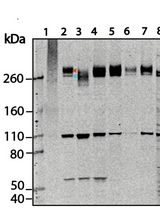
Cathepsin D (catD) is a lysosomal aspartic protease sorted by Mannose 6-phosphate receptors (M6PRs) that transport it from the trans-Golgi network to endosomes/lysosomes in mammalian cells (Ghosh et al., 2003). CatD is synthesized as a precursor protein (~52 kDa), which is cleaved in lysosomes to generate an intermediate (~48 kDa) or the mature lysosomal form (~34 kDa). Trace amounts of the precursor protein are also secreted from the biosynthetic pathway (Benes et al., 2008). The abundance of catD can be determined using several approaches, such as immunofluorescence-based staining (Poole et al., 1972), Western blotting, fluorometric activity assay (Bewley et al., 2011) or metabolic labeling with pulse-chase analysis (Hirst et al., 2009; Kametaka et al., 2007; Tavares et al., 2017). The later is considered a highly sensitive and quantitative approach to monitor catD dynamics (post-translational processing, secretion and degradation) through the secretory pathway. Specifically it involves metabolically labeling newly synthesized proteins in cells followed by a chase, and then to immunoprecipitate catD from the cell lysate and media (secreted form). Therefore, this method provides a way to follow catD molecules from synthesis to lysosomal targeting or secretion with minimal disturbance of normal cell physiology in its natural environment. Herein we described metabolic labeling with [35S] methionine-cysteine in HeLa cells to monitor catD processing, secretion and degradation by using immunoprecipitation, SDS-PAGE and fluorography.
Materials and Reagents
- Filter pipette tips
- 6-well plate (Corning, catalog number: 3516 )
- Ice bucket
- Aluminum foil
- 1.5 ml micro-centrifuge tubes (Corning, Axygen®, catalog number: MCT-150-C )
- WhatmanTM 3030-347 Grade 3 MM Chr cellulose chromatography paper sheet (GE Healthcare, catalog number: 3030-347 )
- Minisart filters pore size 0.22 μm (Sartorius, catalog number: 16534-K )
- HeLa (ATCC, catalog number: CCL-2 )
- 10x PBS (Thermo Fisher Scientific, InvitrogenTM, catalog number: AM9624 )
- Penicillin-streptomycin solution (Thermo Fisher Scientific, GibcoTM, catalog number: 15070063 )
- Fetal bovine serum (FBS) (Thermo Fisher Scientific, catalog number: 12657029 )
- Dulbecco’s modified Eagle’s medium-high glucose-without L-methionine, L-cysteine and L-glutamine (Sigma-Aldrich, catalog number: D0422 )
- L-Methionine (Sigma-Aldrich, catalog number: M9625 )
- L-Cysteine (Sigma-Aldrich, catalog number: 168149 )
- L-Glutamine (Thermo Fisher Scientific, GibcoTM, catalog number: 25030081 )
- EasyTagTM EXPRESS 35S protein labeling mix of both 35S-L-methionine and 35S-L-cysteine (Express Protein Label) (PerkinElmer, catalog number: NEG772007MC )
- Trizma® base (Sigma-Aldrich, catalog number: T1503 )
- Sodium chloride (NaCl) (Sigma-Aldrich, catalog number: S9888 )
- Ethylenediamine tetraacetic acid (EDTA) (Thermo Fisher Scientific, InvitrogenTM, catalog number: 15575020 )
- Triton X-100 (Sigma-Aldrich, catalog number: T8787 )
- Protease inhibitor cocktail (Sigma-Aldrich, catalog number: P8340 )
- Protein A plus Ultralink resin (Thermo Fisher Scientific, catalog number: 53142 )
- 10% (v/v) bovine serum albumin (BSA) (Thermo Fisher Scientific, InvitrogenTM, catalog number: 15561020 )
- Cathepsin D antibody (EMD Millipore, Calbiochem, catalog number: 219361 )
- L-Glutamine (Thermo Fisher Scientific, GibcoTM, catalog number: 25030149 )
- Sodium dodecyl sulfate (SDS) (Sigma-Aldrich, catalog number: L3771 )
- Glycerol (C3H8O3) (Sigma-Aldrich, catalog number: G5516 )
- Bromophenol blue (Sigma-Aldrich, catalog number: B0126 )
- Methanol (CH3OH) (Sigma-Aldrich, catalog number: 322415 )
- Acetic acid (CH3COOH) (Sigma-Aldrich, catalog number: 320099 )
- Amplify fluorographic reagent (GE Healthcare, catalog number: NAMP100 )
- β-Mercaptoethanol (Sigma-Aldrich, catalog number: M3701 )
- Dulbecco’s modified eagle medium (DMEM) (Thermo Fisher Scientific, GibcoTM, catalog number: 12800017 ) (see Recipes)
- 100x L-methionine/L-cysteine solution (see Recipes)
- Pulse medium (see Recipes)
- Chase medium (see Recipes)
- Lysis buffer (see Recipes)
- Wash buffer (see Recipes)
- 2x sample buffer (see Recipes)
- Fixation solution (see Recipes)
- Running buffer (see Recipes)
Equipment
- Pipettes
- Incubator
- Titer plate shaker (Thermo Fisher Scientific, catalog number: 4625Q )
- Centrifuge for 1.5 ml micro-centrifuge tubes (Thermo Fisher Scientific, model: SorvallTM ST 16 , catalog number: 75004380) with rotor for micro-centrifuge tubes (Thermo Fisher Scientific, catalog number: 75003652 )
- Tube rotator capable of end-over-end inversion of the tubes (Phoenix Luferco, catalog number: AP22 or equivalent)
- A chamber to run mini-gels (Bio-Rad Laboratories, model: Mini-PROTEAN Tetra Cell )
- Gel-dryer (Hoefer, model: GD2000 , Slab Gel Dryer) coupled with a vacuum pump
- Exposure cassette for unmounted screen, 20 x 25 cm (GE Healthcare, catalog number: 63-0035-44 )
- Pharos FX plus molecular imager (Bio-Rad Laboratories, catalog number: 1709450 )
- BAS storage phosphor screen (GE Healthcare, catalog number: 28-9564-82 )
- Appropriate receptacle to dispose of solid and liquid contaminated with 35S (according to the local radiation safety guidelines)
Software
- Quantity One 1-D Analysis Software (Bio-Rad Laboratories)
Procedure
文章信息
版权信息
© 2017 The Authors; exclusive licensee Bio-protocol LLC.
如何引用
Tavares, L. A. and daSilva, L. L. (2017). Monitoring the Targeting of Cathepsin D to the Lysosome by Metabolic Labeling and Pulse-chase Analysis. Bio-protocol 7(21): e2598. DOI: 10.21769/BioProtoc.2598.
分类
生物化学 > 蛋白质 > 标记
生物化学 > 蛋白质 > 免疫检测
分子生物学 > 蛋白质 > 检测
您对这篇实验方法有问题吗?
在此处发布您的问题,我们将邀请本文作者来回答。同时,我们会将您的问题发布到Bio-protocol Exchange,以便寻求社区成员的帮助。