- EN - English
- CN - 中文
Nematode Epicuticle Visualisation by PeakForce Tapping Atomic Force Microscopy
通过峰值力轻敲原子力显微镜观察线虫上表皮
发布: 2017年11月05日第7卷第21期 DOI: 10.21769/BioProtoc.2596 浏览次数: 8509
评审: Neelanjan BoseAnonymous reviewer(s)
Abstract
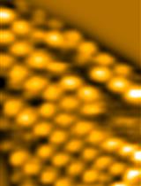
The free-living soil nematode Caenorhabditis elegans has become an iconic experimental model animal in biology. This transparent animal can be easily imaged using optical microscopy to visualise its organs, tissues, single cells and subcellular events. The epicuticle of C. elegans nematodes has been studied at nanoscale using transmission and scanning (SEM) electron microscopies. As a result, imaging artefacts can appear due to embedding the worms into resins or coating the worms with a conductive gold layer. In addition, fixation and contrasting may also damage the cuticle. Conventional tapping mode atomic force microscopy (AFM) can be applied to image the cuticle of the dried nematodes in air, however this approach also suffers from imaging defects. Ideally, the nematodes should be imaged under conditions resembling their natural environment. Recently, we reported the use of PeakForce Tapping AFM mode for the successful visualisation and numerical characterisation of C. elegans nematode cuticle both in air and in liquid (Fakhrullina et al., 2017). We imaged the principal nematode surface structures and characterised mechanical properties of the cuticle. This protocol provides the detailed description of AFM imaging of liquid-immersed C. elegans nematodes using PeakForce Tapping atomic force microscopy.
Keywords: Atomic force microscopy (原子力显微镜检查)Background
Nematodes, both free-living and parasitic, have been extensively studied due to their remarkable biology and effects on agriculture and human well-being. Apparently, the most studied and famous species among nematodes is a free-living soil nematode Caenorhabditis elegans (Sterken et al., 2015). This round worm has been successfully employed as a versatile model organism in a number of investigations (Fire et al., 1998; Kenyon, 2010; Swierczek et al., 2011; O’Reilly et al., 2014; Stroustrup et al., 2016), securing eventually a Nobel prize for Sidney Brenner, John Sulston and Robert Horvitz in 2002. C. elegans nematode is a tiny (~1 mm long) transparent animal which can be easily visualised using an optical microscope. Its cuticle, though thin and transparent, serves as a principal protective barrier between the worm and its habitat. Cuticle is an important marker of disease, when pathogenic bacteria colonize it, leading to the death of the infected animal. In addition, the structure of the cuticle may indicate the ageing in the nematodes. From the human health care and industrial point of view, monitoring of the cuticle of nematodes might be helpful in elucidation of novel antinematode chemicals affecting the cuticle. As a result, nanoscale imaging of the nematode epicuticle in its natural liquid environments may open new avenues in biomedical research.
Previously, the cuticle of nematodes (mostly employing C. elegans as a model organism) has been studied using electron microscopy, both transmission and scanning. Unfortunately, electron microscopy does not allow imaging the worms in liquid, which is their native environment. Sample preparation for electron microscopy requires fixation, dehydration, contrast staining or surface sputtering, and also resin embedding for ultrathin slicing. As a result, nematode cuticle may exhibit secondary artifacts preventing from evaluation of native surface structure and mechanical characterisation. Atomic force microscopy has been established as a potent tool in imaging biological samples in situ, including imaging live cells in liquid media (Beaussart et al., 2015). Recently, the application of tapping mode AFM to visualise C. elegans nematodes in air has been reported (Allen et al., 2015). Imaging in air suffered from the same drawback as scanning electron microscopy, for example the images demonstrated the typical shrunk and collapsed cuticle surfaces apparently caused by dehydration and air imaging. We envisaged a different technique, based on PeakForce Tapping AFM mode (Alsteens et al., 2012) for successful visualisation of C. elegans nematode cuticle in water (Fakhrullina et al., 2017). Here we report a detailed protocol for this technique.
Materials and Reagents
Note: All chemicals were purchased from Sigma-Aldrich unless noted otherwise.
- Pipette tips
- Petri dishes
- Dust-free Nexterion glass slides (Schott)
- Wild type C. elegans (N2 Bristol) nematodes
- Escherichia coli OP50 bacteria
- Poly(allylamine hydrochloride) (PAH, molecular weight ~17,5 kDa) (Sigma-Aldrich, catalog number: 283215 )
- Poly(sodium 4-styrenesulfonate) (PSS, molecular weight ~70 kDa) (Sigma-Aldrich, catalog number: 243051 )
- Nematode growth media (NGM) (bioWORLD, catalog number: 30620040-1 )
- Nematode lysis solution (aqueous 2% NaOCl/0.45 M NaOH)
- Sodium hypochlorite (NaClO) (Sigma-Aldrich, catalog number: 425044 )
- Sodium hydroxide (NaOH) (Sigma-Aldrich, catalog number: S8045 )
- Levamisole hydrochloride (Sigma-Aldrich, catalog number: L9756 )
- Glutaraldehyde (25% aqueous) (Sigma-Aldrich, catalog number: G5882 )
- Ultrapure (type 1) water purified by Simplicity (Millipore) water purification system
- Potassium phosphate monobasic (KH2PO4) (Sigma-Aldrich, catalog number: NIST200B )
- Sodium phosphate dibasic (Na2HPO4) (Sigma-Aldrich, catalog number: 71649 )
- Sodium chloride (NaCl) (Sigma-Aldrich, catalog number: S7653 )
- Magnesium sulfate (MgSO4) (Sigma-Aldrich, catalog number: 793612 )
- Aqueous M9 buffer (see Recipes)
Equipment
- Pipettes (mechanical Eppendorf or Gilson pipettes)
- Eppendorf Scientific Excella E24 temperature-controlled benchtop shaker (Eppendorf, New BrunswickTM, model: Excella® E24 )
- Biosan V-1 plus vortex (Biosan, model: V-1 plus )
- Nikon SMZ 745 T stereomicroscope (Nikon, model: SMZ745T )
- Dimension FastScan Bio atomic force microscope (Bruker, model: Dimension FastScan ) equipped with a liquid submergible scanner
Note: Do not use air scanners for scanning in liquid, this may damage the instrument. - Biosan Microspin 12 centrifuge (Biosan, model: Microspin 12 )
- ScanAsyst-Fluid probes (nominal length 70 µm, tip radius 20 nm, spring constant of 0.7 N m-1) (Bruker, model: SCANASYST-FLUID )
Note: Other AFM probes from Bruker or other producers approved for using in PeakForce Tapping mode having characteristics similar to ScanAsyst-Fluid probes can also be used.
Software
- NanoScope AFM operating software (Bruker Corporation)
- NanoScope Analysis v.1.7. software (Bruker Corporation)
Procedure
文章信息
版权信息
© 2017 The Authors; exclusive licensee Bio-protocol LLC.
如何引用
Akhatova, F., Fakhrullina, G., Gayazova, E. and Fakhrullin, R. (2017). Nematode Epicuticle Visualisation by PeakForce Tapping Atomic Force Microscopy. Bio-protocol 7(21): e2596. DOI: 10.21769/BioProtoc.2596.
分类
细胞生物学 > 细胞成像 > 原子力显微镜
您对这篇实验方法有问题吗?
在此处发布您的问题,我们将邀请本文作者来回答。同时,我们会将您的问题发布到Bio-protocol Exchange,以便寻求社区成员的帮助。
Share
Bluesky
X
Copy link


