

Capturing Z-stacked Confocal Images of Living Bacteria Entering Hydathode Pores of Cauliflower
采集活细菌进入花椰菜泌水孔的Z-栈共聚焦图像

发布: 2017年10月20日第7卷第20期 DOI: 10.21769/BioProtoc.2451 浏览次数: 9217
评审: Anonymous reviewer(s)
参见作者原研究论文
The authors used this protocol in:
Jun 2017
Advertisement


Abstract
The present protocol to visualize living bacteria at the pore level of cauliflower hydathodes is simple and trained users in confocal microscopy can execute it successfully. It can be easily adapted to capture images with other plant-microorganism interactions at the leaf surface and should be useful to obtain important information on pore and stomatal biology. A critical limitation to methods used to observe plant-microorganism interactions in the pore is the application of too much pressure to the sample during observations and z-stack acquisitions. To solve this issue, we recommend the use of a long working-distance water immersion objective lens that allows observations even with thick samples.
Keywords: Cauliflower (花椰菜)Background
Pores of hydathodes and stomata are possible entry points for pathogenic microorganisms to invade plant tissues. In cauliflower, the hydathodes present on leaf margins exhibit large pores, resembling stomata. These pores are routes for the leaf infection by the vascular pathogenic bacterium Xanthomonas campestris pv. campestris (Xcc) (Cerutti et al., 2017). Hydathodes are present on leaves of a wide range of vascular plants. We describe a simple protocol to visualize bacteria at the pore level by confocal microscopy.
Materials and Reagents
- Razor blade
- Microscope glass slides (Thermo Fisher Scientific, Superfrost, catalog number: 10143560WCUT )
- Cover slip 24 x 60 mm (Thermo Fisher Scientific, catalog number: 15747592 )
- Compost (Proveen, catalog number: 14926 )
- Cauliflower (Brassica oleracea var. botrytis, cultivar Clovis, Vilmorin)
Note: Plants were grown on compost in a controlled greenhouse (short day conditions 9 h light; temperature 22 °C; relative humidity 70%). After inoculation, they were placed back in the controlled greenhouse for 24 h inside the miniature greenhouse at 100% relative humidity. After 24 h, lid was removed. All the experiments used the second true leaf from four-week-old plants.
- Xcc strain 8004::GUS-GFP (Xanthomonas campestris pv. campestris (Xcc); Cerutti et al., 2017)
- Magnesium chloride (MgCl2) (Merck, catalog number: 814733 )
- Yeast extract (Sigma-Aldrich, catalog number: Y1625 )
- Casamino acids (BD, BD Biosciences, catalog number: 223050 )
- Potassium phosphate dibasic (K2HPO4) (Merck, catalog number: 105101 )
- Magnesium sulfate heptahydrate (MgSO4·7H2O) (Merck, catalog number: 105886 )
- Silwet L-77 (CAAHMRO, catalog number: 115950H )
- Calcofluor (Fluorescent Brightener 28) (Sigma-Aldrich, catalog number: F3543 )
- MOKA medium (see Recipes)
Equipment
- Miniature greenhouse purchased from a garden center (Nortene, catalog number: ME673714 )
- Hollow punch (Harris, Uni-Core) (Electron Microscopy Sciences, catalog number: 69039-70 ) or razor blades
- Confocal microscope (Leica Microsystems, model: Leica TCS SP2 or Leica TCS SP8 ) equipped with an argon laser (ray line at 488 nm), a Helium Neon laser at 633 nm and a diode laser at 405 nm
- Water immersion lens 40x with a long working distance (N.A. = 0.8, working distance of 3,300 µm) (Leica Microsystems, catalog number: 50615 5) or water immersion lens 63x with a long working distance (N.A. = 0.9 and a working distance of 2,900 µm) (Leica Microsystems, catalog number: 506148 ) (Leica Mannheim, Germany)
Procedure
文章信息
版权信息
© 2017 The Authors; exclusive licensee Bio-protocol LLC.
如何引用
Readers should cite both the Bio-protocol article and the original research article where this protocol was used:
- Cerutti, A. and Jauneau, A. (2017). Capturing Z-stacked Confocal Images of Living Bacteria Entering Hydathode Pores of Cauliflower. Bio-protocol 7(20): e2451. DOI: 10.21769/BioProtoc.2451.
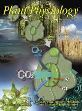
- Cerutti, A., Jauneau, A., Auriac, M. C., Lauber, E., Martinez, Y., Chiarenza, S., Leonhardt, N., Berthomé, R. and Noël, L. D. (2017). Immunity at cauliflower hydathodes controls systemic infection by Xanthomonas campestris pv campestris. Plant Physiol 174(2): 700-716.
分类
植物科学 > 植物细胞生物学 > 细胞成像
微生物学 > 微生物-宿主相互作用 > 体内实验模型 > 植物
细胞生物学 > 细胞成像 > 共聚焦显微镜
您对这篇实验方法有问题吗?
在此处发布您的问题,我们将邀请本文作者来回答。同时,我们会将您的问题发布到Bio-protocol Exchange,以便寻求社区成员的帮助。