

Isolation and Analysis of Stromal Vascular Cells from Visceral Adipose Tissue
.内脏脂肪组织来源基质血管细胞的分离和分析
发布: 2017年08月20日第7卷第16期 DOI: 10.21769/BioProtoc.2444 浏览次数: 13946
评审: Jia LiAnonymous reviewer(s)
参见作者原研究论文
The authors used this protocol in:
Mar 2017
Advertisement


Abstract
The obesity epidemic is the underlying driver of the type 2 diabetes mellitus epidemic. A remarkable accumulation of various pro-inflammatory immune cells in adipose tissues is a hallmark of obesity and leads to pathogenesis of tissue inflammation and insulin resistance. Here, we describe a detailed protocol to isolate adipose tissue stromal vascular cells (SVCs), which enrich various immune cells of adipose tissues. These SVCs can be used to examine the population and activation status of immune cells by tracking their cell surface antigens, gene expression, and activation of specific signaling pathways.
Keywords: Adipose tissue (脂肪组织)Background
Over the past several decades, obesity is now an epidemic and has become one of the most common causes of insulin resistance. Insulin resistance is the key etiology for the pathogenesis of metabolic syndrome. Prolonged status of metabolic syndrome drives the development of type 2 diabetes mellitus (T2DM) (Romeo et al., 2012; Johnson and Olefsky, 2013; Saltiel and Olefsky, 2017).
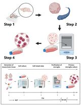
Chronic low-degree tissue inflammation, accompanied by enhanced immune cell infiltration, is a hallmark of obesity in both rodent and human and is a major causal factor for the pathogenesis of insulin resistance through promoting the inflammation status and interrupting the insulin signalling (Romeo et al., 2012; Johnson and Olefsky, 2013; Saltiel and Olefsky, 2017). The infiltrated immune cells such as pro-inflammatory macrophages and B cells play critical roles in modulating obesity-associated adipose tissue inflammation and insulin resistance (Weisberg et al., 2003; Winer et al., 2011). Chronic nutrient excess drives adipose tissue macrophages (ATMs) to undergo a unique phenotypic switch from anti-inflammatory M2-like activation in lean adipose tissue to a more pro-inflammatory M1-like activation state in obese tissues (Lumeng et al., 2007; Nguyen et al., 2007; Lumeng et al., 2008). Pro-inflammatory M1-like ATMs contribute to the development of tissue inflammation and systemic insulin resistance in obesity. Our recent study also demonstrates that leukotriene B4 (LTB4)-induced recruitment and activation of adipose tissue B2 (ATB2) cells can cause obesity-induced insulin resistance (Ying et al., 2017). In this protocol, we provide a step-by-step procedure to isolate stromal vascular cells from adipose tissue and characterize various immune cells in adipose tissues.
Materials and Reagents
- Pipette tips (USA Scientific)
- 100-mm Petri dish
- 50 ml Falcon tube (Corning, Falcon®, catalog number: 352070 )
- Nylon biopsy bag (Electron Microscopy Sciences, catalog number: 62324-35 )
- MicroAmp Optical 96-well reaction plate (Thermo Fisher Scientific, Applied BiosystemsTM, catalog number: N8010560 )
- Stromal vascular cells (SVCs)
- 70% ethanol
- BDTM stabilizing fixative buffer (BD, BD Biosciences, catalog number: 339860 )
- Phosphate-buffered saline (PBS)
- 2% fetal bovine serum (FBS)
- Antibody
- Rabbit monoclonal anti-GAPDH (Cell Signaling Technology, catalog number: 5174 )
- Rabbit monoclonal anti-Phospho-NF-κB p65 (Cell Signaling Technology, catalog number: 3033 )
- PE-Cyanine7 anti-mouse F4/80 (Thermo Fisher Scientific, eBioscienceTM, catalog number: 25-4801-82 )
- Alexa Fluor 488 anti-mouse CD11b (Thermo Fisher Scientific, eBioscience TM, catalog number: 53-0112-82 )
- APC anti-mouse CD11c (Thermo Fisher Scientific, eBioscienceTM , catalog number: 17-0114-82 )
- PE anti-mouse CD206 (BioLegend, catalog number: 141706 )
- eVolve-605 anti-mouse CD45 (Thermo Fisher Scientific, eBioscienceTM, catalog number: 83-0451-42 )
- APC anti-mouse CD19 (Thermo Fisher Scientific, eBioscienceTM , catalog number: 17-0193-82 )
- Rabbit monoclonal anti-GAPDH (Cell Signaling Technology, catalog number: 5174 )
- Trizol reagent (Thermo Fisher Scientific, InvitrogenTM , catalog number: 15596026 )
- Direct-zol RNA kits (Zymo Research, catalog number: R2070 )
- High-Capacity cDNA Reverse Transcription Kit (Thermo Fisher Scientific, Applied BiosystemsTM, catalog number: 4368813 )
- qPCR primers (Table 1)
Table 1. qPCR primer information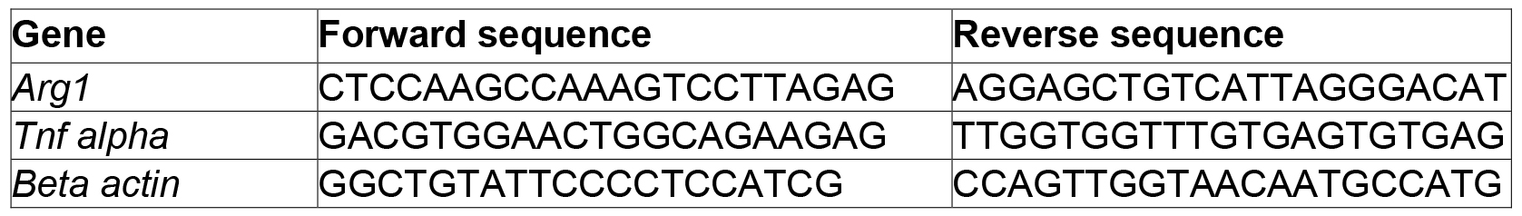
- SYBR Green PCR Master mix (Thermo Fisher Scientific, Applied BiosystemsTM, catalog number: 4309155 )
- Hanks’ balanced salt solution (HEPES) (Thermo Fisher Scientific, GibcoTM, catalog number: 15630080 )
- Collagenase II (Sigma-Aldrich, catalog number: C1764-50MG )
- Bovine serum albumin (BSA)
- Ammonium chloride (NH4Cl)
- Potassium bicarbonate (KHCO3)
- 5% EDTA
- Sodium azide (NaN3)
- Iscove’s Modified Dulbecco’s Medium (IMDM)
- Penicillin-streptomycin (Thermo Fisher Scientific, GibcoTM, catalog number. 15140122 )
- Digestion buffer (see Recipes)
- Red blood cell lysis buffer (see Recipes)
- FACS staining buffer (see Recipes)
- Complete culture medium (see Recipes)
Equipment
- Pipettes
- Mortar and pestle
- Curved scissors
- New Brunswick Scientific 12400 incubator shaker (Eppendorf, model: New BrunswickTM 124 )
- Eppendorf centrifuge 5810R (Eppendorf, model: 5810 R )
- TC20 automated cell counter (Bio-Rad Laboratories, model: TC20TM, catalog number: 1450102 )
- BD FACSCanto flow cytometry analyzer
- StepOnePlus Real-Time PCR System (Thermo Fisher Scientific, Applied BiosystemsTM, model: StepOnePlusTM , catalog number: 4376600)
- DNA Engine Peltier Thermal Cycler (Bio-Rad Laboratories, model: PTC-200 )
- NanoDrop 1000 Spectrophotometer (Thermo Fisher Scientific, model: NanoDropTM 1000 )
Software
- FlowJo
- GraphPad Prism
Procedure
文章信息
版权信息
© 2017 The Authors; exclusive licensee Bio-protocol LLC.
如何引用
Vu, J. and Ying, W. (2017). Isolation and Analysis of Stromal Vascular Cells from Visceral Adipose Tissue. Bio-protocol 7(16): e2444. DOI: 10.21769/BioProtoc.2444.
分类
免疫学 > 免疫细胞分离 > 基质血管细胞
细胞生物学 > 细胞分离和培养 > 细胞分离
您对这篇实验方法有问题吗?
在此处发布您的问题,我们将邀请本文作者来回答。同时,我们会将您的问题发布到Bio-protocol Exchange,以便寻求社区成员的帮助。