

[2-3H]Mannose-labeling and Analysis of N-linked Oligosaccharides
N-连接寡糖的[2-3H] 甘露糖标记和分析

发布: 2017年07月20日第7卷第14期 DOI: 10.21769/BioProtoc.2393 浏览次数: 7708
评审: Ralph BottcherAnonymous reviewer(s)
参见作者原研究论文
The authors used this protocol in:
Aug 2016


Abstract
Modifications of N-linked oligosaccharides of glycoproteins soon after their biosynthesis correlate to glycoprotein folding status. These alterations can be detected in a sensitive way by pulse-chase analysis of [2-3H]mannose-labeled glycoproteins, with enzymatic removal of labeled N-glycans, separation according to size by HPLC and radioactive detection in a scintillation counter.
Keywords: N-linked oligosaccharide (N-连接寡糖)Background
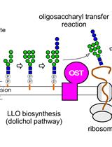
Following entry of a nascent polypeptide into the ER, it is subjected to several post-translational modifications, which are crucial for folding, maturation and quality control processes. The addition of the core oligosaccharide, Glc3Man9GlcNAc2 to produce N-linked glycoproteins is a very common modification and the first to occur (Benyair et al., 2011). Processing of the precursor N-glycan directs the glycoproteins to maturation and quality control machineries by creating recognition tags while in early secretory compartments (Tannous et al., 2015). At later stages, throughout the secretory pathway the core of the oligosaccharide serves as a platform for expansion of the sugar chains into complex glycans, the structures of which relate to the trafficking and function of the glycoproteins (Kamiya et al., 2012). Because the early N-linked glycan modifications reflect glycoprotein biosynthesis and quality control, the oligosaccharide processing has been the subject of many studies (Avezov et al., 2010; Hosokawa et al., 2010; Ninagawa et al., 2014; Ogen-Shtern et al., 2016). Glycoproteomic methods have greatly improved N-glycan characterization, but they do not allow the study of the dynamics of glycan processing in the early secretory pathway.
Here we describe a simplified pulse-chase method for the isolation and analysis of metabolically labeled N-linked oligosaccharides. The method includes radioactive labeling by [2-3H]Man followed by enzymatic removal of oligosaccharides by endo-beta-N-acetylglucosaminidase H (Endo H). Then, N-linked oligosaccharide isolation by molecular filtration and separation by high-performance liquid chromatography (HPLC), which discriminates between high-mannose glycan structures depending on their number of monosaccharide residues. The protocol allows analysis of the dynamics of N-linked glycan modification under different conditions, e.g., after drug treatment or modification of protein levels by overexpression or knockdown. Trimming to shorter species, Man5-6GlcNAc2, is a requirement for glycoprotein targeting to endoplasmic reticulum-associated degradation (ERAD) (Frenkel et al., 2003). Man1A (α1,2 mannosidase 1A ) appears to be involved in endoplasmic reticulum (ER) quality control and required for this trimming, as we present in an example. This is a surprising finding considering that the enzyme was thought to be located in the Golgi complex (Igdoura et al., 1999; Herscovics, 2001); a recent reevaluation locates it in quality control vesicles (Ogen-Shtern et al., 2016).
To conclude, the protocol presented here enables the study of the dynamics of N-linked high-mannose glycan modifications, which has an important role in glycoprotein quality control and trafficking. The advantages of the method are its simplicity, high sensitivity of detection and unique information on the dynamics of N-glycan processing in the early secretory pathway.
Materials and Reagents
- 100 mm tissue culture dishes (Corning, catalog number: 430167 )
- 1.5 and 2 ml microcentrifuge tubes (Eppendorf)
- Microcon Amicon Ultra 0.5 ml 30K or Centricon ultracel YM-30 (EMD Millipore, catalog number: UFC503024 )
- Spherisorb NH2 column, 5 µm, 4.6 x 250 mm (WATERS, catalog number: PSS831115 )
- HEK-293 cells (ATCC, catalog number: CRL-1573 )
- Dulbecco’s modified Eagle’s medium (DMEM) (Thermo Fisher Scientific, GibcoTM, catalog number: 41965039 )
- Dulbecco’s modified Eagle’s medium-glucose free (Sigma-Aldrich, catalog number: D5030 )
- Fetal bovine serum (FBS) (Biological Industries, catalog number: 04-001-1A-US )
- Fetal bovine serum (FBS), dialyzed (Biological Industries, catalog number: 04-011-1A-US )
- Sodium pyruvate solution (100 mM) (Sigma-Aldrich, catalog number: S8636 )
- Mannose, D-[2-3H(N)]- (Specific Activity: 15-30 Ci/mmol) (PerkinElmer, catalog number: NET570A )
- Dulbecco’s phosphate buffered saline (PBS) (Sigma-Aldrich, catalog number: D1408 )
- Protein A-Agarose beads, IPA 300 (REPLIGEN, catalog number: 10-1003 )
- Rabbit polyclonal anti-H2 carboxy-terminal produced in our lab (Tolchinsky et al., 1996)
- Endo Hf Kit (1,000,000 U/ml) (New England Biolabs, catalog number: P0703S )
- Standard oligosaccharide mixture prepared by glycoprotein metabolic labeling with [14C] or [3H] and separation with endo H
- Acetonitrile LiChrosolv (gradient grade for liquid chromatography) (Merck, catalog number: 100030 )
- Phosphoric acid solution (49-51%, for HPLC) (Sigma-Aldrich, catalog number: 79607 )
- Opti-Fluor scintillation fluid (PerkinElmer, catalog number: 6013199 )
- Triton X-100 (BDH, catalog number: 306324N )
- Protease inhibitor cocktail (Sigma-Aldrich, catalog number: P2714 )
- Sodium deoxycholate (Sigma-Aldrich, catalog number: 30970 )
- Sodium dodecyl sulfate (SDS) (Bio-Rad Laboratories, catalog number: 1610301 )
- Sodium phosphate (Na3PO4) (Sigma-Aldrich, catalog number: 342483 )
- Buffer A (see Recipes)
- Buffer D (see Recipes)
- HPLC solvent (see Recipes)
Equipment
- Eppendorf centrifuge (Eppendorf, model: 5415 D )
- CO2 incubator
- -80 °C freezer
- LS 6500 Liquid Scintillation Counting systems (Beckman Coulter, model: LS 6500, catalog number: 510720 )
- 600E Multisolvent delivery System controller (HPLC) (WATERS, catalog number: WAT062710 )
Note: This product has been discontinued. - SpeedVac Concentrator 5301, incl. 48 x 1.5/2.0 ml fixed-angle rotor (Eppendorf, model: Concentrator 5301, catalog number: 5301 000.016 )
- Frac-100 fraction collector (Amersham Biosciences, model: FRAC-100, catalog number: 18-1000-77 )
- Vibra-Cell ultrasonic processors VCX 750 (Sonics and Materials, model: VCX 750 , catalog number: 690-003)
Procedure
文章信息
版权信息
© 2017 The Authors; exclusive licensee Bio-protocol LLC.
如何引用
Shenkman, M., Ogen-Shtern, N. and Lederkremer, G. Z. (2017). [2-3H]Mannose-labeling and Analysis of N-linked Oligosaccharides. Bio-protocol 7(14): e2393. DOI: 10.21769/BioProtoc.2393.
分类
生物化学 > 糖类 > 寡糖 > 标记
您对这篇实验方法有问题吗?
在此处发布您的问题,我们将邀请本文作者来回答。同时,我们会将您的问题发布到Bio-protocol Exchange,以便寻求社区成员的帮助。
![]() 提问指南
提问指南
+ 问题描述
写下详细的问题描述,包括所有有助于他人回答您问题的信息(例如实验过程、条件和相关图像等)。