

Endpoint or Kinetic Measurement of Hydrogen Sulfide Production Capacity in Tissue Extracts
组织提取物中生成硫化氢的终点或动力学测定
发布: 2017年07月05日第7卷第13期 DOI: 10.21769/BioProtoc.2382 浏览次数: 11528
评审: Neelanjan BoseTanxi CaiAnonymous reviewer(s)
参见作者原研究论文
The authors used this protocol in:

Jan 2015
Advertisement


Abstract
Hydrogen sulfide (H2S) gas is produced in cells and tissues via various enzymatic processes. H2S is an important signaling molecule in numerous biological processes, and deficiencies in endogenous H2S production are linked to cardiovascular and other health complications. Quantitation of steady-state H2S levels is challenging due to volatility of the gas and the need for specialized equipment. However, the capacity of an organ or tissue extract to produce H2S under optimized reaction conditions can be measured by a number of current assays that vary in sensitivity, specificity and throughput capacity. We developed a rapid, inexpensive, specific and relatively high-throughput method for quantitative detection of H2S production capacity from biological tissues. H2S released into the head space above a biological sample reacts with lead acetate to form lead sulfide, which is measured on a continuous basis using a plate reader or as an endpoint assay.
Keywords: Hydrogen sulfide production capacity (硫化氢生成能力)Background
Hydrogen sulfide (H2S) gas is produced endogenously by at least three different enzymes in mammals (CGL, CBS, 3-MST) with a range of tissue and cell-type distributions. H2S functions as a gasotransmitter and effector molecule (Wang, 2012) in a wide range of biological functions related to metabolism (Módis et al., 2013), stress resistance (Hine et al., 2015), and redox biology (Dickhout et al., 2012). Reduced H2S is linked to cardiovascular problems including hypertension in rodents (Yang et al., 2008) and cardiac hypertrophy in man (Polhemus et al., 2014). Increased H2S can also cause pathology, for example in rodent pancreatitis (Bhatia et al., 2005). Thus, accurate and quantitative detection of H2S from biological sources could facilitate a better understanding of its biological effects as well as its potential use as a clinical biomarker.
Techniques to measure absolute concentrations of H2S present in biological samples, along with their pros and cons, have been reviewed extensively (Olson, 2012; Wang, 2012; Hartle and Pluth, 2016; Takano et al., 2016). For example, free and sulfane-bound H2S pools can be measured in biological samples including serum or tissue homogenates ex vivo using headspace GC-MS, which is highly sensitive and selective, but requires expensive equipment. Nonetheless, due to the volatility of H2S, its interaction with other biological macromolecules and its breakdown into different sulfur-containing compounds, quantitative detection of steady-state free H2S levels in vivo remains challenging (Olson, 2009).
An alternate approach is to measure the capacity of a tissue homogenate or extract to produce H2S in a reaction mixture containing optimized levels of substrate and cofactor, thus allowing for H2S detection methods that are specific but less sensitive. An example is the methylene blue method in which H2S in solution is trapped by lead acetate to form lead sulfide, which upon conversion to methylene blue can be easily read in a standard spectrophotometer (Stipanuk and Beck, 1982; Ikeda et al., 2017). The pros and cons that must be taken into account with each method are based on the question being asked, the biological system and tissue being studied, the relative need for sensitivity, selectivity, or speed, and the cost and resources of the investigator.
Here, we describe an inexpensive, rapid, and moderately high throughput methodology for measuring H2S production capacity in extracts of relatively small amounts of biological material. This method is based on the reaction of H2S present in the headspace above a biological sample with lead acetate to form the black precipitate lead sulfide, a technique used throughout the past 100 years to detect H2S and H2S-producing bacteria (McBride and Edwards, 1914; Kuester and Williams, 1964; Zhang and Weiner, 2014). Previously, we used this method to detect changes in H2S production capacity as a function of diet or genetic background in a variety of biological samples including yeast, worms, flies, and rodent tissues/organs including liver (Hine et al., 2015; Mitchell et al., 2016; Nikonorova et al., 2017). Here, we present an optimized procedure to measure H2S production capacity in mammalian liver via (B) an end-point assay using Whatman paper-embedded lead acetate, or (C) a kinetic assay using agar-embedded lead acetate. As the liver is a strong producer of H2S in mammalian systems via the enzyme cystathionine gamma lyase (CGL) (Kabil et al., 2011), we feel this is a good starting point for researchers to understand and confidently develop this protocol for their own research questions. Furthermore, this procedure can be easily adapted to other biological samples and organisms, although the procedure may need to be optimized by the investigator in order to obtain suitable results.
Materials and Reagents
- Petri dish
- 1.5 ml RNase-free disposable pellet pestles and 1.5 ml tubes (Fisher Scientific, catalog number: 12-141-368 )
- Disposable razor blades
- 8-strip well format tubes (Denville Scientific)
- Hard/rigid plastic dissecting platform/sheet
- Plastic wrap
- Filter paper (703 Style Whatman)
- 96-well plates with lid (Corning, catalog number: 3370 )
- Gloves and proper personal protective equipment
- 15 ml centrifuge tube
- Flash frozen mouse livers
- De-ionized water
- Phosphate buffered saline (PBS), pH 7.4 (Fisher Scientific, catalog number: BP24384 )
- Liquid nitrogen
- Ice
- Dry ice
- 5x passive lysis buffer (Promega, catalog number: E1941 )
- BCA Protein Assay Kit (Thermo Fisher Scientific, Thermo ScientificTM, catalog number: 23227 )
- L-cysteine (Sigma-Aldrich, catalog number: C7352 )
- Pyridoxal 5’-phosphate hydrate (Sigma-Aldrich, catalog number: P9255 )
- Lead(II) acetate trihydrate (Sigma-Aldrich, catalog number: 316512 )
- Agarose (HS Molecular Biology Grade) (Denville Scientific, catalog number: CA3510-8 , or use similar)
Note: This product has been discontinued. - 1x passive lysis buffer (see Recipes)
- 20 mM lead(II) acetate trihydrate (see Recipes)
- H2S reaction mixture (see Recipes)
- 1% agarose gel with 100 mM lead(II) acetate trihydrate (see Recipes)
Equipment
- Liquid nitrogen flask (Thermo flask 2122) (Thermo Fisher Scientific, Thermo ScientificTM, catalog number: 2122 )
- Forceps
- Scale (OHAUS, catalog number: EP214C )
- Pipettes and tips (single use and multichannel for pipetting between 2 µl to 5 ml)
- -80 °C freezer
- Motorized tissue grinder (Fisher Scientific, catalog number: 12-1413-61 )
- 37 °C water bath
- Micro-centrifuge (VWR, model: Galaxy 16DH )
- UV-Vis plate reader (BioTek Instruments, model: Synergy 2 )
- Large glass Pyrex baking dish (> 100 ml)
- Glass flask (> 100 ml capacity)
- Vacuum oven (VWR, catalog number: 89508-424 )
- Incubator (VWR, model: 1500E )
- Digital camera (Kodak, model: KODAK EASYSHARE C182 )
- Vortex mixer (Scientific Industries, model: Vortex-Genie 2 , catalog number: SI-0236)
- Computers (HP Pavilion dv6 and Lenovo IdeaPad)
- Heat block cube (9.5 x 7.5 x 5 cm), or other heavy object with approximate dimensions
Software
- GraphPad Prism 7
- Microsoft Excel
- ImageJ
- Gen5
Procedure
文章信息
版权信息
© 2017 The Authors; exclusive licensee Bio-protocol LLC.
如何引用
Hine, C. and Mitchell, J. R. (2017). Endpoint or Kinetic Measurement of Hydrogen Sulfide Production Capacity in Tissue Extracts. Bio-protocol 7(13): e2382. DOI: 10.21769/BioProtoc.2382.
分类
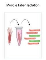
细胞生物学 > 组织分析 > 组织分离
细胞生物学 > 细胞信号传导 > 第二信使
生物化学 > 其它化合物 > 硫化氢
您对这篇实验方法有问题吗?
在此处发布您的问题,我们将邀请本文作者来回答。同时,我们会将您的问题发布到Bio-protocol Exchange,以便寻求社区成员的帮助。