

Ex vivo Ooplasmic Extract from Developing Drosophila Oocytes for Quantitative TIRF Microscopy Analysis
从在发育的果蝇卵母细胞中离体提取卵磷脂用于定量TIRF显微镜分析


发布: 2017年07月05日第7卷第13期 DOI: 10.21769/BioProtoc.2380 浏览次数: 9444
评审: Jihyun KimAnonymous reviewer(s)
参见作者原研究论文
The authors used this protocol in:
Feb 2017
Advertisement


Abstract
Understanding the dynamic behavior and the continuously changing composition of macromolecular complexes, subcellular structures and organelles is one of areas of active research in both cell and developmental biology, as these changes directly relate to function and subsequently to the development and homeostasis of the organism. Here, we demonstrate the use of the developing Drosophila oocyte to study dynamics of messenger ribonucleoprotein complexes (mRNPs) with high spatiotemporal resolution. The combination of Drosophila genetics with total internal reflection (TIRF) microscopy, image processing and data analysis gives insight into mRNP motility and composition dynamics with unprecedented precision.
Keywords: Ooplasmic extract (卵磷脂提取)Background
Intracellular transport is one of the fundamental processes in living cells. Almost everything within the cell–ions, molecules, complexes, organelles–is transported actively such that the local entropy is reduced. Although in recent years we have gained considerable understanding of the mechanisms underlying these transport process, most of our knowledge comes from in vitro and cell culture studies. In these simplified systems, it is difficult to establish whether the full potential of the transport regulatory processes is utilized. Tissues, organs, organoids and organisms, on the other hand, are often too complex to be studied efficiently with spatiotemporal resolution sufficient to match the scale of these transport processes. To combine the advantages of the bottom-up and top-down approaches, techniques have been developed that, while preserving complexity, make these processes more accessible. One example is the preparation of mass cytoplasmic extract from ambiphian (e.g., Xenopus laevis) oocytes and embryos to study cell divisions (Lohka and Masui, 1983; Murray, 1991; Sawin and Mitchison, 1991). We have recently shown that in cytoplasmic drops–i.e., non-purified cytoplasm directly extracted from the cell–released from single Drosophila embryos mitotic activity of the contained nuclei continues, allowing the probing of spindle properties by simple physical and chemical perturbations (Telley et al., 2012 and 2013). Here, we describe a similar ex vivo preparation technique based on ooplasm of developing Drosophila egg-chambers. This method allows the study of intracellular transport processes (squash assay), such as the transport of localizing oskar mRNPs (Gaspar et al., 2017).
Materials and Reagents
- Coverslip stand for 5-10 coverslips (e.g., Wash-N-Dry, Diversified Biotech, catalog number: WSDR-1000 )
- Gloves
- A small plastic Petri dish or the cap of a 50 ml Falcon tube (~30 mm diameter)
- 15 x 20 cm black plastic plate
- High precision coverslips (e.g., Marienfeld Precision Cover Glass, 22 x 22 mm, Marienfeld-Superior, catalog number: 0107052 )
- X-ray film (e.g., Amersham HyperfilmTM ECL, GE Healthcare, catalog number: 28906835 )
- Double-sided adhesive tape (e.g., Tesa Doubleband Photostrip, Tesa, catalog number: 05338-00 )
- Cheesecloth
- A fly line or a cross that yields females of the appropriate genotype (see Note 1)
- Dry, granular baker’s yeast (e.g., Lesaffre Saf-Instant®)
- Halocarbon oil (e.g., Voltalef 10S, VWR, catalog number: 24627.188 )
- 100% ethanol (e.g., EMD Millipore, catalog number: 100983 )
- ~5% dichlorodimethylsilane (DCDMS) in heptane (e.g., Silanization Solution, Sigma-Aldrich, catalog number: 85126 )
- PIPES (e.g., Sigma-Aldrich, catalog number: P3768 )
- Magnesium chloride hexahydrate (MgCl2·6H2O) (e.g., EMD Millipore, catalog number: 105833 )
- EGTA (e.g., Sigma-Aldrich, catalog number: E3889 )
- HEPES (e.g., Sigma-Aldrich, catalog number: H3375 )
- Potassium chloride (KCl) (e.g., EMD Millipore, catalog number: 104936 )
- Dextran sulfate, MW ~10 kDa (e.g., Sigma-Aldrich, catalog number: D4911 )
- Agar (e.g., from Pro-BIO)
- Dry yeast (e.g., from Volk Klaus)
- Soya powder (e.g., from Ruckemann)
- Sirup (e.g., from Ruckemann)
- Malt extract (e.g., from Baeko Rhei)
- Corn powder (e.g., from Ruckemann)
- Propionic acid (e.g., VWR, catalog number: ACRO447231000)
Manufacturer: Acros Organics, catalog number: 447231000 . - Nipagin (e.g., Sigma-Aldrich, catalog number: H5501 )
- BRB80 (see Recipes)
- 1% injection buffer (1% IB) (see Recipes)
- Cornmeal agar (see Recipes)
Equipment
- Standard fly husbandry equipment (e.g., vials with cornmeal agar–see Recipes, 25 °C incubator with humidity controller, brushes, CO2 station, etc.)
- 3-9 well dissection plate (e.g., Corning, catalog number: 7220-85 )
- Vacuum desiccator (e.g., SP Scienceware - Bel-Art Products - H-B Instrument, catalog number: F42025-0000 )
- Dumont #5 forceps (e.g., Fine Science Tools, catalog number: 11252-20 )
- Dumont #55 forceps (e.g., Fine Science Tools, catalog number: 11255-20 )
- Custom made coverslip holder for microscopy (Figure 1)
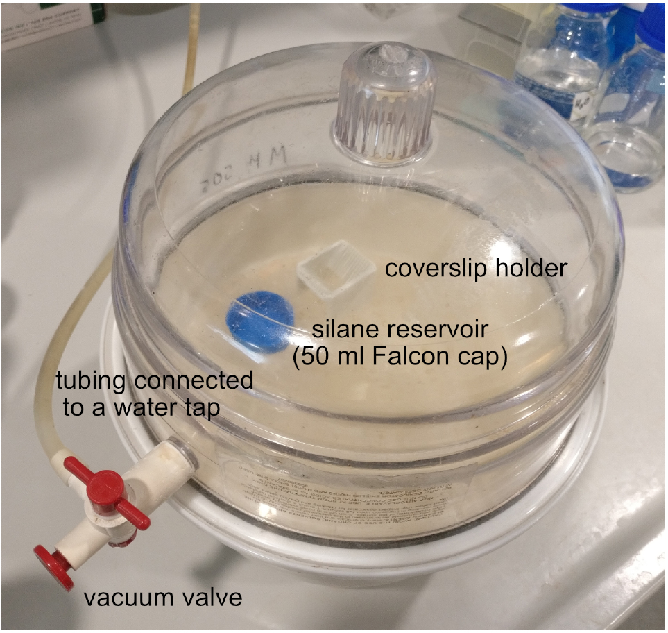
Figure 1. Vacuum desiccator loaded with the coverslip holder and the silane reservoir. After replacing the lid of the device, apply vacuum by opening the vacuum valve and the water tap. Once the vacuum is formed–i.e., you can lift the entire device by the lid–close the vacuum valve and the water tap. - Zooming stereomicroscope with 5-40x (or higher) magnification (e.g., Carl Zeiss, model: STEMI SV 11 )
- Sharp tungsten needles with handles (e.g., Fine Science Tools, catalog numbers: 10130-20 and 26018-17 )
- Total Internal Reflection Fluorescent (TIRF) microscope equipped with a high NA objective and a high sensitivity detector (Leica Microsystems, model: Leica AF7000 )
Note: We use a Leica 7000 wide-field TIRF microscope with a 100x 1.46 NA oil immersion objective and a Photometrics Evolve Roper 512 EMCCD camera.
Software
- Microscope controller software (e.g., Leica LASAF)
- ImageJ/FIJI
- Custom-made particle detector and tracker plug-in for ImageJ (available to download under https://github.com/Xaft/xs/blob/master/_xs.jar)
- R (preferentially with RStudio), for data analysis
Procedure
文章信息
版权信息
© 2017 The Authors; exclusive licensee Bio-protocol LLC.
如何引用
Gáspár, I. and Ephrussi, A. (2017). Ex vivo Ooplasmic Extract from Developing Drosophila Oocytes for Quantitative TIRF Microscopy Analysis. Bio-protocol 7(13): e2380. DOI: 10.21769/BioProtoc.2380.
分类
发育生物学 > 细胞信号传导 > 配体
细胞生物学 > 细胞成像 > 荧光
您对这篇实验方法有问题吗?
在此处发布您的问题,我们将邀请本文作者来回答。同时,我们会将您的问题发布到Bio-protocol Exchange,以便寻求社区成员的帮助。