

Photothrombotic Induction of Capillary Ischemia in the Mouse Cortex during in vivo Two-Photon Imaging
在体内双光子成像期间采用光栓法诱导小鼠皮层毛细血管缺血
发布: 2017年07月05日第7卷第13期 DOI: 10.21769/BioProtoc.2378 浏览次数: 10388
评审: Pengpeng LiZinan ZhouAnonymous reviewer(s)
参见作者原研究论文
The authors used this protocol in:

Nov 2016
Advertisement


Abstract
Photothrombosis of blood vessels refers to the activation of a circulating photosensitive dye with a green light to induce clotting in vivo (Watson et al., 1985). Previous studies have described how a focused green laser could be used to noninvasively occlude pial arterioles and venules at the brain surface (Schaffer et al., 2006; Nishimura et al., 2007; Shih et al., 2013). Here we show that small regions of the capillary bed can similarly be occluded to study the ischemic response within the capillary system of the mouse cerebral cortex. The advantage of this approach is that the ischemic zone is restricted to a diameter of approximately 150-250 μm. This permits higher quality two-photon imaging of degenerative processes that would be otherwise difficult to visualize with models of large-scale stroke, due to excessive photon scattering. A consequence of capillary occlusion is leakage of the blood-brain barrier (BBB). Here, through the use of two-photon imaging data sets, we show how to quantify capillary leakage by determining the spatial extent and localization of intravenous dye extravasation.
Keywords: Blood-brain barrier (血脑屏障)Background
Numerous animal models exist to induce ischemia on a large scale via occlusion major cerebral arteries (Carmichael, 2005). However, there are some aspects of stroke that are not accessible to in vivo two-photon imaging. In regions experiencing more severe ischemia, cells swell due to ionic imbalance, and this edematous process contributes to increased light scattering, greatly reducing the quality and depth of in vivo two-photon imaging. A smaller zone of ischemia would reduce photon scattering, and still allow neurovascular changes associated with ischemia to be more clearly visualized over time in vivo.
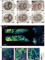
We recently showed that spatially restricted regions of ischemia could be generated by focused photothrombotic irradiation of the cortical capillary bed (Underly et al., 2017). Capillary occlusions were highly reproducible, could be targeted to specific locations, and initiated at precise times through a cranial imaging window. The resulting ischemic zone occupied less than 1% of the area accessible through a typically cranial window (Figures 1D and 1E), allowing multiple strokes to be examined in one window.
Here, we describe the steps involved in inducing photothrombotic occlusion in a small region of the capillary bed during in vivo two-photon imaging. We build upon a previous protocol in which individual cortical penetrating arterioles were occluded, rather than capillaries (Taylor and Shih, 2013). We also demonstrate the analysis of BBB leakage produced as a consequence of capillary occlusion using Imaris, a 3-D visualization software.
Materials and Reagents
- Filter paper (GE Healthcare, catalog number: 1001-0155 )
- Cotton swabs (Fisher Scientific, catalog number: 23-400-119 )
- Cover glass (thickness: No. 0) (Thomas Scientific, catalog number: 6661B40 )
- 0.3 ml insulin syringes (BD, catalog number: 328438 )
- Petri dish
- An adult mouse of any common laboratory strain, ~25 to 35 g in weight
- Isoflurane (Patterson Veterinary Supply, catalog number: 07-806-3204 )
Manufacturer: Zoetis, catalog number: 10015516 . - Phosphate buffered saline (PBS) (Sigma-Aldrich, catalog number: P4417-50TAB )
- Agarose type 3-A (Sigma-Aldrich, catalog number: A9793 )
- Fluorescein-dextran (2 MDa; 5% w/v in saline) (Sigma-Aldrich, catalog number: FD2000S )
- Rose Bengal (1.25% w/v in saline) (Sigma-Aldrich, catalog number: 330000-1G )
- Buprenorphine hydrochloride (Buprenex®) (Patterson Veterinary Supply, catalog number: 07-891-9756 )
- Sodium chloride (NaCl) (Sigma-Aldrich, catalog number: S7653-1KG )
- Potassium chloride (KCl) (Sigma-Aldrich, catalog number: P9333-500G )
- Calcium chloride (CaCl2) (Sigma-Aldrich, catalog number: C1016-100G )
- Magnesium chloride (MgCl2) (Sigma-Aldrich, catalog number: M8266-100G )
- Glucose (Sigma-Aldrich, catalog number: G8270 )
- HEPES (Sigma-Aldrich, catalog number: H7006 )
- Artificial cerebral spinal fluid (ACSF) (see Recipes)
Equipment
- Heating pad with feedback regulation (FHC, catalog number: 40-90-2-07 )
- Heating pad control system (FHC, catalog number: 40-90-8D )
- Rectal Thermistor Probe (FHC, catalog number: 40-90-5D-02 )
- Isoflurane vaporizer (Datex-Ohmeda, model: IsoTec4 )
- Induction chamber (VetEquip, catalog number: 941444 )
- Dental drill (Osada, model: EXL-M40 )
- Auxiliary equipment for two-photon microscope (Sutter Moveable Objective System [Taylor and Shih, 2013])
- Objective lens 4x, 0.16 NA (Olympus, model: UPLSAPO )
- Objective lens 20x, 1.0 NA, Water immersion (Olympus, model: XLUMPlanFI )
- Green laser 532 nm (Beta Electronics, model: MGM20 ). Details for how the green laser line is directed into the Sutter MOM imaging beam path was described in a separate protocol (Taylor and Shih, 2013)
- Computer specifications (for Imaris)
- 16 GB RAM
- 3.3 GHz CPU
- AMD Radeon RX 480 4GB or better
- 1280 x 1024 Resolution Monitor
Software
- Imaris (Bitplane)
- Imaris 7.6 (or current)
- Imaris Batch Module
- Fiji software (https://imagej.net/Fiji/Downloads)
Procedure
文章信息
版权信息
© 2017 The Authors; exclusive licensee Bio-protocol LLC.
如何引用
Underly, R. G. and Shih, A. Y. (2017). Photothrombotic Induction of Capillary Ischemia in the Mouse Cortex during in vivo Two-Photon Imaging. Bio-protocol 7(13): e2378. DOI: 10.21769/BioProtoc.2378.
分类
细胞生物学 > 细胞成像 > 双光子显微镜
您对这篇实验方法有问题吗?
在此处发布您的问题,我们将邀请本文作者来回答。同时,我们会将您的问题发布到Bio-protocol Exchange,以便寻求社区成员的帮助。