

Qualitative and Quantitative Assay for Detection of Circulating Autoantibodies against Human Aortic Antigen
人主动脉抗原循环自身抗体的定性和定量检测实验

发布: 2017年07月05日第7卷第13期 DOI: 10.21769/BioProtoc.2367 浏览次数: 10461
评审: Jia LiPorkodi PanneerselvamAnonymous reviewer(s)
参见作者原研究论文
The authors used this protocol in:
July 2015
Advertisement


Abstract
Increased amount of autoantibodies in human sera are the hallmark of autoimmune diseases (Wang et al., 2015). In case of known antigen, detection of autoantibodies is done using laboratory based methods. However, in most autoimmune diseases, knowledge of self-antigen is still vague. We have developed an ELISA-based quantitative assay to detect the presence of autoantibodies as well as to measure the circulating autoantibodies in the sera of patients suffering from large vessel vasculitis (LVV), an autoimmune disease (Chakravarti et al., 2015). Using this assay, we detected the increase in anti-aortic antibodies in LVV patient’s sera. We have further verified the results by independent biochemical techniques and found the specificity to be > 94% (Chakravarti et al., 2015). This method can be uniquely modified to suit any autoimmune, in particular organ specific, disease and thus has wider applications in the detection and quantification of autoantibodies.
Keywords: ELISA (ELISA)Background
There are about 80-100 autoimmune diseases wherein immune cells recognize a self-protein as a foreign antigen, and get activated to generate humoral response. Though the trigger for most autoimmune diseases remains a mystery, presence of higher levels of antibodies in the patient’s sera is very common (Wang et al., 2015). Some of the examples include antibodies against neutrophil cytoplasmic antigens in small vessel vasculitis, myelin basic protein in multiple sclerosis, glutamate decarboxylase in type 1 diabetes etc. (Wang et al., 2015). Limited knowledge of autoantigen in most autoimmune diseases presents a challenge for both the detection of disease as well as understanding the pathogenesis. However, ability to detect autoantibodies in the sera provides a diagnostic and prognostic advantage. Discovering a reliable autoantigen in autoimmune diseases are needed to make better clinical decisions. LVV is a spectrum of autoimmune diseases affecting aorta or its primary branch vessels. Like many others, its etiology and cure are not known (Buttgereit et al., 2016). We have developed a qualitative and quantitative assay to detect the presence of autoantibody in the human sera targeting against the human aortic antigen. Our assay provides a tool to find autoantigen in any affected/damaged tissue and in any disease model. We looked for the presence of autoantigen in the human thoracic aortic aneurysms using patients’ sera and tested the specificity of the assay by comparing the sera obtained from related subsets of autoimmune or non-immune diseases. We utilized discarded aortic tissues from aortic reconstruction surgeries and made soluble extracts of aorta to set up the assay (Figure 1). Using this method we performed a high throughput screen for detecting autoantibody in more than 100 sera against aortic antigen (Chakravarti et al., 2015).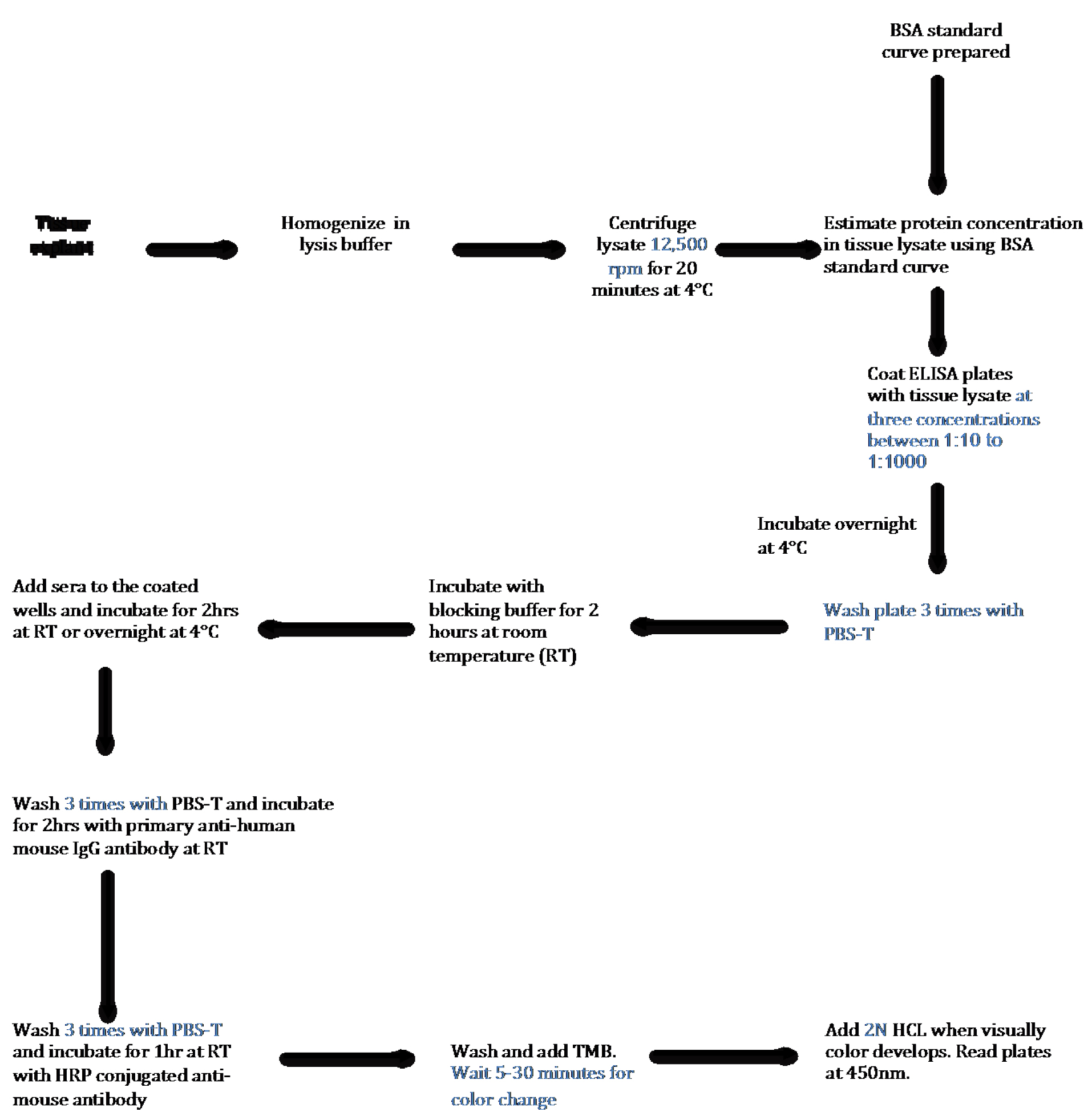
Figure 1. Schematic Flow Diagram of ELISA to detect autoantibody in human tissue lysates. Step-by-step outline of the method used to detect and quantify autoantibodies present in the sera against antigen present in tissue.
Materials and Reagents
- 5 ml sterile polystyrene round bottom tubes (Corning, Falcon®, catalog number: 352008 )
- 1.5 ml Eppendorf tubes (USA Scientific, catalog number: 1615-5500 )
- 15 ml centrifuge tubes (Thermo Fisher Scientific, Thermo ScientificTM, catalog number: 339650 )
- Greiner 96 well, F-Bottom, clear microplate (Greiner Bio One International, catalog number: 655001 )
- 200 μl pipette tip (USA Scientific, catalog number: 1111-1700 )
- 1,250 μl pipette tip (USA Scientific, catalog number: 1112-1720 )
- Immulon 2 HB: high affinity protein binding plates (Thermo Fisher Scientific, Thermo ScientificTM, catalog number: 3455 )
- Paper towel
- Human Specimen
Note: Human tissue explant as well as sera should be collected under institutionally approved IRB protocol. - Protease
- Phosphatase inhibitor (PhosSTOP) (Roche Diagnostics, catalog number: 04906837001 )
- Protein Assay dye (Bio-Rad Laboratories, catalog number: 5000006 )
- Bovine serum albumin (BSA) (Sigma-Aldrich, catalog number: A9418 )
- Phosphate-buffered saline-Tween (PBS-T) (Fisher Scientific, catalog number: BP293810 )
- Antibodies:
- Ultra-TMB (Thermo Fisher Scientific, Thermo ScientificTM, catalog number: 34029 )
- 2 N HCl (Fisher Scientific, catalog number: SA431-500 )
- Tris buffer, pH 7.4 (Fischer Scientific, catalog number: BP152-1 )
- Sodium chloride (NaCl) (Sigma-Aldrich, product number: S9888 )
- Triton X-100 (Bio-Rad Laboratories, catalog number: 1610407 )
- Sodium orthovanadate (Sigma-Aldrich, catalog number: S6508 )
- Sodium fluoride (Sigma-Aldrich, catalog number: 919 )
- Glycerophosphate (Sigma-Aldrich, catalog number: G9422 )
- Sodium pyrophosphate (Sigma-Aldrich, catalog number: P8010 )
- Non-fat dry milk powder (Bio-Rad Laboratories, catalog number: 1706404 )
- Cell lysis buffer (see Recipes)
- Blocking buffer (see Recipes)
Equipment
- 2-20 μl pipettes (Alkali Scientific, catalog number: P9280-20U )
- 20-200 μl pipettes (Alkali Scientific, catalog number: P9280-200U )
- Scissors (Fisher Scientific, catalog number: 08-951-20 )
- Table top centrifuge (Labnet International, model: PrismRTM R, catalog number: C2500-R )
- Orbital shaker (Benchmark Scientific, model: BT302 )
- Refrigerator
- Plate reader (BMG LABTECH, model: FLUstar Omega )
- Homogenizer (Thomas Scientific, model: SCILOGEX D160 )
Software
- MS Excel
- GraphPad Prism 7
Procedure
文章信息
版权信息
© 2017 The Authors; exclusive licensee Bio-protocol LLC.
如何引用
Veerman, B. and Chakravarti, R. (2017). Qualitative and Quantitative Assay for Detection of Circulating Autoantibodies against Human Aortic Antigen. Bio-protocol 7(13): e2367. DOI: 10.21769/BioProtoc.2367.
分类
免疫学 > 抗体分析 > 抗体检测
生物化学 > 蛋白质 > 定量
您对这篇实验方法有问题吗?
在此处发布您的问题,我们将邀请本文作者来回答。同时,我们会将您的问题发布到Bio-protocol Exchange,以便寻求社区成员的帮助。