

An Acute Mouse Spinal Cord Slice Preparation for Studying Glial Activation ex vivo
用于研究胶质细胞离体激活的急性小鼠脊髓切片的制备


发布: 2017年01月20日第7卷第2期 DOI: 10.21769/BioProtoc.2102 浏览次数: 11943
评审: Oneil G. BhalalaJingli CaoKae-Jiun Chang
参见作者原研究论文
The authors used this protocol in:
Apr 2016


Abstract
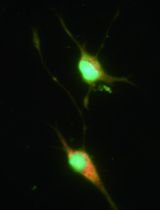
Pathological conditions such as amyotrophic lateral sclerosis, spinal cord injury and chronic pain are characterized by activation of astrocytes and microglia in spinal cord and have been modeled in rodents. In vivo imaging at cellular level in these animal models is limited due to the spinal cord’s highly myelinated funiculi. The preparation of acute slices may offer an alternative and valuable strategy to collect structural and functional information in vitro from dorsal, lateral and ventral regions of spinal cord. Here, we describe a procedure for preparing acute slices from mouse spinal cord (Garré et al., 2016). This preparation should allow further understanding of how glial cells in spinal cord respond acutely to various inflammatory challenges.
Keywords: Microglia (小胶质细胞)Background
Mouse transgenic technology has been used to model different human pathologies affecting the spinal cord, many of which are characterized by local glial activation, one hallmark of neuroinflammation. A major breakthrough that has enormously increased the understanding of glial biology in health and disease is the utilization of laser scanning microscopy based techniques, such as confocal microscopy (White et al., 1987) and two-photon microscopy (Denk et al., 1990) to visualize cell structures and subcellular domains in living animals in a noninvasive fashion; for example, mice expressing genetically encoded reporters or calcium sensors have been used to image glial structures (somata and processes) and to study calcium dynamics and signaling, respectively (Davalos et al., 2005; Gee et al., 2014). In spinal cord, myelin is highly compact in the white matter of the dorsal, lateral, and ventral funiculi. In vivo structural imaging of glial cells and infiltrating immune cells has been successfully performed in the past using surgical procedures (laminectomy) that allow optical access to the dorsal spinal cord (Kim et al., 2010). However, since myelin greatly increases light scattering, imaging is limited to the superficial layers of the dorsal funiculus, masking valuable information from deeper regions such as ventral horn. We think that acute slices prepared from wild type and transgenic mice can be used in combination with high-resolution imaging techniques to offer an alternative strategy to collect structural and functional information, in vitro, from dorsal, and also lateral and ventral regions. Coronal sections interrupt ascending and descending axons and many motor axons as well. Nevertheless, the information obtained is likely to be useful in analyzing how glial cells respond acutely to inflammatory challenges in spinal cord.
Materials and Reagents
- Double edge razor blades (Everychina, Baili, catalog number: BP005 )
- Sterile 21 gauge needles (BD, catalog number: 305165 )
- Syringes (½ ml, 3 ml, and 20 ml)
½ ml (COVIDIEN, catalog number: 8881600004 )
3 ml (BD, catalog number: 309657 )
20 ml (BD, catalog number: 302830 ) - Adhesive tape
- Peel-a-way embedding molds (Sigma-Aldrich, catalog number: E6032 )
- Disposable transfer pipettes (Thermo Fisher Scientific, Thermo ScientificTM, catalog number: 336 )
- Six-well multidish (Thermo Fisher Scientific, catalog number: 130184 )
- Parafilm
- Coverslip (Thermo Fisher Scientific, Fisher Scientific, catalog number: 12-545-88 )
- 70 μm cell strainer (Corning, Falcon®, catalog number: 352350 )
- 15 ml and 50 ml polypropylene conical tubes (Corning, Falcon®, catalog numbers: 352095 and 352098 , respectively)
- Pipette tips (10 μl, 200 μl, 1,000 μl) (USA Scientific)
- One- to two-month-old CX3CR1EGFP/+ transgenic mice (THE JACKSON LABORATORY, catalog number: 005582 )
- Ketamine and xylazine (provided by NYU School of Medicine, DLAR)
- Isoflurane (provided by NYU School of Medicine, DLAR)
- 70% ethanol
- Low melting point agarose (Sigma-Aldrich, catalog number: A9414 )
- Cyanoacrylate (Instant Krazy glue)
- Ethidium bromide (MW: 394.3) (MP Biomedicals, catalog number: 802511 )
- Propidium iodide (MW: 668.4) (Thermo Fisher Scientific, Molecular ProbesTM, catalog number: P3566 )
- Phosphate buffered saline (Thermo Fisher Scientific, GibcoTM, catalog number: 14190-144 )
- Triton X-100, 100 ml solution (Sigma-Aldrich, catalog number: X100 )
- Bovine serum albumin (BSA) (Sigma-Aldrich, catalog number: A3294 )
- Normal goat serum (Vector Laboratories, catalog number: S1000 )
- Optional: chicken anti-GFAP (EMD Millipore, catalog number: AB5541 )
- Mowiol® 4-88 (aqueous mounting medium) (Sigma-Aldrich, catalog number: 81381 )
- Tween 20
- Optional: alexa fluor 647-conjugate goat anti-chicken IgY - H&L (Thermo Fisher Scientific, Invitrogen, catalog number: A21449 ) secondary antibody
- Sodium chloride (NaCl) (Sigma-Aldrich, catalog number: S7653 )
- Sodium bicarbonate (NaHCO3) (Sigma-Aldrich, catalog number: S5761 )
- Glucose (Sigma-Aldrich, catalog number: G7528 )
- Potassium chloride (KCl) (Sigma-Aldrich, catalog number: P9333 )
- Sodium phosphate monobasic (NaH2PO4) (Sigma-Aldrich, catalog number: S8282 )
- Calcium chloride (CaCl2) (Sigma-Aldrich, catalog number: C1016 )
- Magnesium chloride (MgCl2) (Sigma-Aldrich, catalog number: M8266 )
- HCl
- NaOH
- EDTA
- Paraformaldehyde (PFA, 32% solution) (Electron Microscopy Sciences, catalog number: 15714 )
- Artificial cerebrospinal fluid (ACSF) (see Recipes)
- Ca2+ and Mg2+ free-ACSF (see Recipes)
- 3-4% PFA, pH = 7.4 (see Recipes)
Equipment
- Compressed gas tank 5% CO2, 95% O2
- Leica vibratome and blade holder (Leica, model: VT1000 S )
- Standard 1000 orbital shaker (TROEMNER, catalog number: 980173 )
- Digital pH meter (Mettler Toledo)
- Hemostat clamps (World Precision Instruments, catalog number: 503736 )
- Forceps 12 cm long (World Precision Instruments, catalog number: 14226 )
- Fine dissection forceps number 5 (Roboz Surgical Instrument, catalog number: RS-4955 )
- SuperCut scissors (World Precision Instruments, catalog number: 14218 )
- Spine bone scissors (Dumont, catalog number: 15a )
- Digital water bath (Thermo Fisher Scientific, Fisher ScientificTM, model: Isotemp 205 )
- Tubing
- Digital scale (Mettler Toledo, model: MS104S )
- Micropipettes (Gilson, 0.5-2 µl, 1-10 µl , 10-200 µl , 1,000 µl )
- Stereo microscope with LED lights (Olympus, model: SZX10 )
- Zeiss-700 confocal microscope equipped with 20x objective and appropriate filters
- Thermistor thermometer (SP Scienceware - Bel-Art Products - H-B Instrument, catalog number: 605010100 )
Procedure
文章信息
版权信息
© 2017 The Authors; exclusive licensee Bio-protocol LLC.
如何引用
Readers should cite both the Bio-protocol article and the original research article where this protocol was used:
- Garré, J. M., Yang, G., Bukauskas, F. F. and Bennett, M. V. L. (2017). An Acute Mouse Spinal Cord Slice Preparation for Studying Glial Activation ex vivo. Bio-protocol 7(2): e2102. DOI: 10.21769/BioProtoc.2102.
-
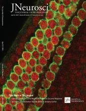
Garré, J. M., Yang, G., Bukauskas, F. F. and Bennett, M. V. (2016). FGF-1 triggers Pannexin-1 hemichannel opening in spinal astrocytes of rodents and promotes inflammatory responses in acute spinal cord slices. J Neurosci 36(17): 4785-4801.
分类
神经科学 > 细胞机理 > 细胞分离和培养
细胞生物学 > 细胞分离和培养 > 细胞分离
您对这篇实验方法有问题吗?
在此处发布您的问题,我们将邀请本文作者来回答。同时,我们会将您的问题发布到Bio-protocol Exchange,以便寻求社区成员的帮助。