- EN - English
- CN - 中文
Generation of Tumour-stroma Minispheroids for Drug Efficacy Testing
用于检测药物疗效的肿瘤基质小球体的生成
发布: 2017年01月05日第7卷第1期 DOI: 10.21769/BioProtoc.2091 浏览次数: 9991
评审: HongLok LungKevin Patrick O’RourkeVikash Verma

相关实验方案

基于Fiji ImageJ的全自动化流程开发:批量分析共聚焦图像数据并量化蛋白共定位的Manders系数
Vikram Aditya [...] Wei Yue
2025年04月05日 2841 阅读
Abstract
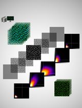
The three-dimensional organisation of cells in a tissue and their interaction with adjacent cells and extracellular matrix is a key determinant of cellular responses, including how tumour cells respond to stress conditions or therapeutic drugs (Elliott and Yuan, 2011). In vivo, tumour cells are embedded in a stroma formed primarily by fibroblasts that produce an extracellular matrix and enwoven with blood vessels. The 3D mixed cell type spheroid model described here incorporates these key features of the tissue microenvironment that in vivo tumours exist in; namely the three-dimensional organisation, the most abundant stromal cell types (fibroblasts and endothelial cells), and extracellular matrix. This method combined with confocal microscopy can be a powerful tool to carry out drug sensitivity, angiogenesis and cell migration/invasion assays of different tumour types.
Keywords: Mixed cell type 3-dimensional (3D) culture (混合细胞类型三维(3D)培养)Background
The traditional monolayer cell culture (2-dimensional) enforces an artificial environment, which is vastly different from the tissues cells exists in vivo. One of the most critical differences is that in monolayer cultures the cells are polarised, i.e., the surface of the cells facing the culture-plastic and the upper cell surface exposed to the culture medium receive completely different, often opposing signals (Fitzgerald et al., 2015). To address the problem of cell polarization, tumour spheroid cultures are increasingly used in cancer research. Tumour spheroids can replicate the 3-dimensional cell-cell interactions present in a tissue and to some extent paracrine signaling via cytokines and chemokines by reducing their diffusion and dilution by the growth medium that typically occurs in monolayer cultures (Lawlor et al., 2002; Barrera-Rodríguez and Fuentes, 2015). The current tumour-stroma minispheroid protocol is one such method. Compared to the other tumour-spheroid protocols, this method also incorporates additional, key features of the tissue environment, namely stromal cells and extracellular matrix in the spheroid and thus provides a model that replicates the in vivo tumour microenvironment more faithfully.
Materials and Reagents
- 96 U-shaped well plate for suspension cells (Greiner Bio One, catalog number: 650161 )
- FiltopurTM syringe filters (SARSTEDT, catalog number: 83.1826.001 )
- 50 ml syringes (TERUMO, catalog number: SS+50ES )
- 12 well dishes with 10 mm diameter glass bottom (MATTEK, catalog number: P12G-0-10-F )
- 1.5 ml sterile Eppendorf tubes (SARSTEDT, catalog number: 72.690.001 )
- 50 ml sterile centrifuge tubes (Corning, catalog number: 430829 )
- 35 mm glass bottom dish, 14 mm diameter (MATTEK, catalog number: P35G-0.170-14-C )
- T75 flasks for adherent cells (SARSTEDT, catalog number: 83.3911 )
- Serological pipettes (5 ml, 10 ml) (CORNING, catalog numbers: 4051 and 4101 , respectively)
- Pipette Tips (10 μl, 200 μl, 1,000 μl) (SARSTEDT, catalog numbers: 70.1130.100 , 70.760.002 and 70.762.100 , respectively)
- Cell lines: MDA-MB-231 breast cancer epithelial cells (ATCC, HTB-26TM, catalog number: MDA-MB-231); human umbilical vein endothelial cells (HUVEC) (ATCC, CRL-1730TM, catalog number: HUV-EC-C ); normal human dermal fibroblasts (NHDF) (Lonza, catalog number: CC-2509 )
- Recombinant human tumour necrosis factor-related apoptosis-inducing ligand (rhTRAIL) (purified in-house), receptor-selective TRAIL mutant, TRAIL-45 (O’Leary et al., 2016; van der Sloot et al., 2006)
- Dulbecco’s modified Eagle medium (DMEM)-low glucose concentration (Sigma-Aldrich, catalog number: D6046 )
- Fetal bovine serum (Sigma-Aldrich, catalog number: F7524 )
- L-glutamine solution, 200 Mm stock (Sigma-Aldrich, catalog number: G7513 )
- 1x trypsin-EDTA buffer in HBSS
- CellTrackerTM CM-Dil Dye (Thermo Fisher Scientific, Molecular ProbesTM, catalog number: C7001 ) or CMTPX red cell tracker dye (Thermo Fisher Scientific, Molecular ProbesTM, catalog number: C34552 )
- Rat tail collagen type I (Corning, catalog number: 354236 )
- Hoechst33342 – 10 mg/ml solution in water (Thermo Fisher Scientific, Molecular ProbesTM, catalog number: H3570 )
- SYTOX Green nucleic acid dye (Thermo Fisher Scientific, Molecular ProbesTM, catalog number: S7020 )
- Endothelial cell growth medium-2 (EGM-2) prepared by adding EGMTM-2 SingleQuotsTM Kit (Lonza, catalog number: CC-4176 ) to EBM-2 basal Medium (Lonza, catalog number: CC-3156 )
- Hanks’ balanced salt solution (HBSS) (Thermo Fisher Scientific, GibcoTM catalog number: 24020117 )
- 1 N NaOH solution
- Methylcellulose solution (see Recipes)
Equipment
- HeraeusTM MegafugeTM centrifuge (15 ml, 50 ml tube) (Thermo Fisher Scientific, Thermo ScientificTM, model: 16 Centrifuge Series )
- Mammalian cell culture incubator (37 °C, 5% CO2) (Thermo Fisher Scientific, Thermo ScientificTM, model: FormaTM Steri-CycleTM )
- Hemocytometer
- Pipettes (10 μl, 200 μl, 1,000 μl)
- Pipette aid
- Confocal microscopy system (AndorTM, Revolution Spinning Disk Confocal systemTM)
- High-resolution EMCCD camera (Andor iXon EM+)
- Olympus IX81 motorised inverted microscope, fitted with a variable temperature/CO2 humidified incubation chamber for live cell experiments
- Yokagawa CSU22 spinning disk confocal unit
- Magnetic stirrer
- Orbital shaker
Software
- VolocityTM software (PerkinElmer)
Procedure
文章信息
版权信息
© 2017 The Authors; exclusive licensee Bio-protocol LLC.
如何引用
Watters, M. and Szegezdi, E. (2017). Generation of Tumour-stroma Minispheroids for Drug Efficacy Testing. Bio-protocol 7(1): e2091. DOI: 10.21769/BioProtoc.2091.
分类
癌症生物学 > 通用技术 > 肿瘤微环境 > 细胞外基质
细胞生物学 > 细胞成像 > 共聚焦显微镜
您对这篇实验方法有问题吗?
在此处发布您的问题,我们将邀请本文作者来回答。同时,我们会将您的问题发布到Bio-protocol Exchange,以便寻求社区成员的帮助。
Share
Bluesky
X
Copy link


