

Quantitative 3D Time Lapse Imaging of Muscle Progenitors in Skeletal Muscle of Live Mice
活小鼠骨骼肌中肌肉祖细胞的定量三维延时成像


发布: 2016年12月20日第6卷第24期 DOI: 10.21769/BioProtoc.2066 浏览次数: 11207
评审: Antoine de MorreeXiaoyi ZhengAnonymous reviewer(s)
参见作者原研究论文
The authors used this protocol in:
Feb 2016


Abstract
For non-optically clear mammalian tissues, it is now possible to use multi-photon microscopy to penetrate deep into the tissue and obtain detailed single cell images in a live animal, i.e., intravital imaging. This technique is in principle applicable to any fluorescently marked cell, and we have employed it to observe stem cells during the regenerative process. Stem cell-mediated skeletal muscle regeneration in the mouse model has been classically studied at specific time points by sacrificing the animal and harvesting the muscle tissue for downstream analyses. A method for direct visualization of muscle stem cells to gain real-time information over a long period in a live mammal has been lacking. Here we describe a step-by-step protocol adapted from Webster et al. (2016) to quantitatively measure the behaviors of fluorescently labeled (GFP, EYFP) muscle stem and progenitor cells during homeostasis as well as following muscle injury.
Keywords: Muscle stem cell (肌肉干细胞)Background
Long-term in vivo imaging of stem and progenitor cells was first used for hair follicles during continuous physiological regeneration without surgical procedure (Rompolas et al., 2012). By contrast, stem cells for skeletal muscles are largely quiescent and inactive during the normal homeostatic state. An injury to the muscle is necessary to activate muscle stem cells to mount a regenerative process. In vitro live imaging of muscle stem/progenitor cells has been widely used to study them in artificial settings. To understand muscle stem cell behavior during regeneration in their native environment, we developed a method to image them during skeletal muscle regeneration. Our method allows up to 8 h of continuous imaging per session daily following injury. This is the first time that skeletal muscle stem cells have been observed in vivo in an injured/regenerative environment (Webster et al., 2016).
Materials and Reagents
- Lab tape (VWR, catalog number: 89097 )
- Razor blades for shaving hair (VWR, catalog number: 55411-060 )
- Kimwipes (ULINE)
- Transfer pipette (Globe Scientific, catalog number: 137038 )
- Cyanoacrylate based adhesive that bonds skin to glass (i.e., Loctite glass glue; Amazon.com; Scognamiglio et al., 2016)
- Mouse with GFP or YFP expression in Pax7-expressing muscle stem cells (or cell types of interest); GFP and YFP Cre-reporter mice and Pax7-Cre-ERT2 mouse strains are available at Jackson Laboratory (THE JACKSON LABORATORY, catalog numbers: 021847 , 006148, and 012476, respectively)
- Isoflurane (Patterson Veterinary Supply, IsoFlo®, catalog number: 07-806-3204 ; To be applied directly into the vapor chamber of equipment list 4; indicated by arrow in Figure 2)
- EtOH, 70% (Decon Labs, catalog number: V1401 )
- Distilled water (in house)
Equipment
- An inverted Leica SP5 (or equivalent) equipped with a Leica 25x/0.95 water objective, a 35 mm culture dish holder attached to the stage, and nondescanned detectors with a dichroic mirror separating the detectable spectrum (430-550 nm) at 495 nm (Figure 1)
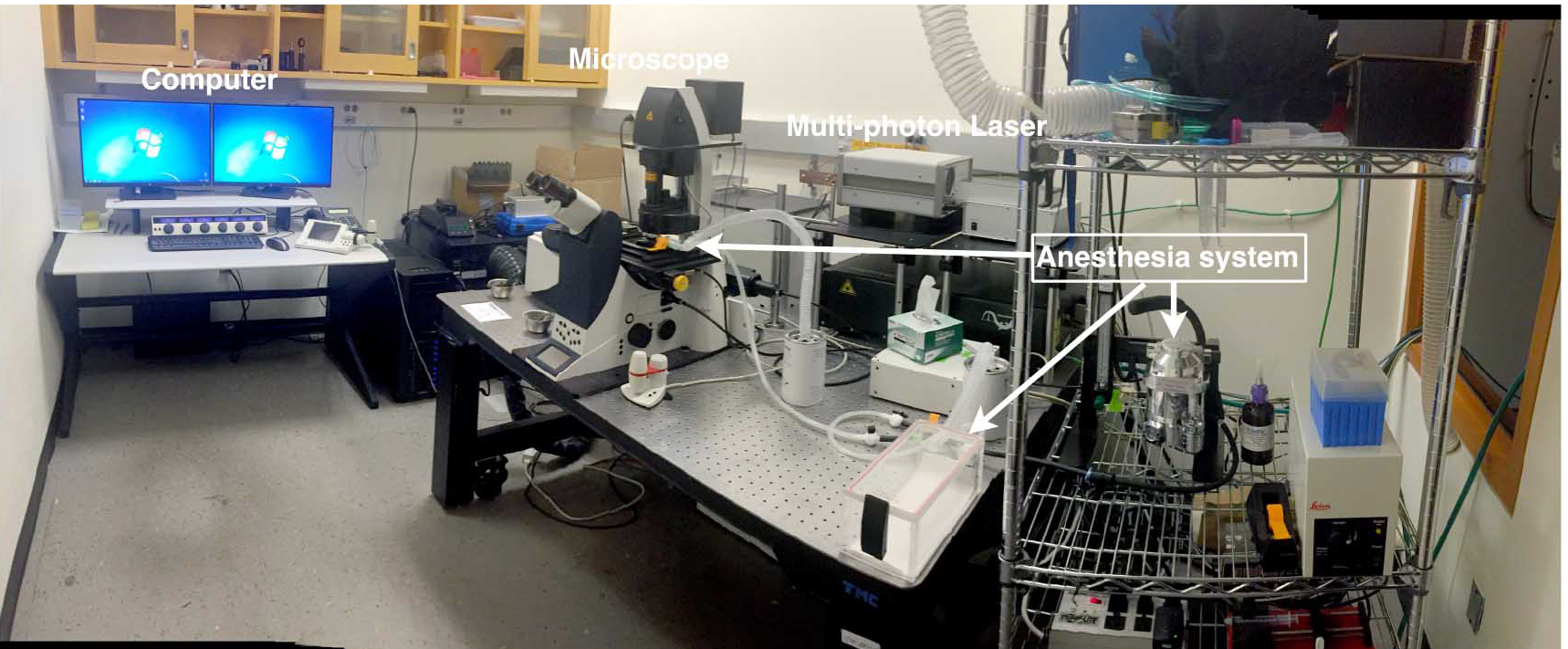
Figure 1. Inverted Multi-photon setup. The imaging setup includes an inverted confocal microscope with 25x water objective fitted with a culture dish holder, multi-photon laser and computer. - Chameleon Vision laser (Coherent)
- Heating pad with enough area to cover the animal on the microscope stage (Sunbeam 756-500 Heating Pad from Amazon.com)
- Animal anesthesia system equipped with induction chamber as well as tubing with nose-cone (VetEquip, catalog number: 901806 ). Please consult with the operation manual prior to use
- 35 mm fluorodish (World Precision Instruments, catalog number: FD35-100 )
- Metal spatula (VWR, catalog number: 82027-530 )
- Heat protective glove (VWR)
- Bunsen burner (VWR, catalog number: 89038-528 )
- Laminar flow fume hood
- Fine tip forceps (Dumont #5 forceps) (Fine Science Tools, catalog number: 11252-30 )
- Fine scissors for cutting skin (straight, 11.5 cm) (Fine Science Tools, catalog number: 14058-11 )
- Spring scissors for cutting fascia (8 mm blades) (Fine Science Tools, catalog number: 15009-08 )
Software
- Leica SP5 software
- Fiji (Fiji is an updated version of ImageJ, an open source image processing software; Schindelin et al., 2012)
- Imaris (Bitplane, version 7.6.4 for Windows X64)
Procedure
文章信息
版权信息
© 2016 The Authors; exclusive licensee Bio-protocol LLC.
如何引用
Webster, M. T., Harvey, T. and Fan, C. (2016). Quantitative 3D Time Lapse Imaging of Muscle Progenitors in Skeletal Muscle of Live Mice. Bio-protocol 6(24): e2066. DOI: 10.21769/BioProtoc.2066.
分类
干细胞 > 成体干细胞 > 肌肉干细胞
细胞生物学 > 细胞成像 > 活细胞成像
您对这篇实验方法有问题吗?
在此处发布您的问题,我们将邀请本文作者来回答。同时,我们会将您的问题发布到Bio-protocol Exchange,以便寻求社区成员的帮助。
![]() 提问指南
提问指南
+ 问题描述
写下详细的问题描述,包括所有有助于他人回答您问题的信息(例如实验过程、条件和相关图像等)。